- Home
- About the Journal
- Peer Review
- Editorial Board
- For Authors
- Reviewer Recognition
- Archive
- Contact
- Impressum
- EWG e.V.
Cite as: Archiv EuroMedica. 2022. 12; 6: e1. DOI 10.35630/2022/12/6.19
The frequency of complications associated with the use of cosmetic invasive interventions to rejuvenate and correct the shape of various parts of the face requires a deep study of the causes of keloid scarring induction. The paper presents an analysis of clinical material in the age aspect, depending on gender and topography of skin lesions. It was noted that in men, injuries are more often associated with burn injuries of the extremities. It has been established that complications of facial skin regeneration are more often observed in women aged 35 to 65 years, often against the background of the use of low-quality invasive materials and in patients with a history of keloid scars.
Keywords: epidermis, skin, proliferation, regeneration, scar, alteration, skin rejuvenation, fibrosis.
According to WHO analytical data, about 50 million people are injured annually in the world; more than 100 million undergo surgery, accompanied by keloid and hypertrophic scars, which is 1.5-4.5% of the total number of operated patients. At the same time, the structure of complications lacks an exhaustive explanation of the causes of interruption of skin regeneration and the formation of keloid scars and the development, in some cases, of keloid disease [4]. Matsumoto N.M., Peng W.X., Aoki M., Akaishi S., Ohashi R., Ogawa R., Naito Z. (2017) the reasons for only the growth of keloid scars and the absence of their regression are associated with a lack of understanding at the present stage of the mechanism of their formation [8]. Many studies have shown the important role of genetic factors in the formation of keloids [3]. Li X., Chen Z., Li X., Wang H. (2019) pointed out that tumor necrosis factor-stimulated gene-6 (TSG-6) plays a key role in the progression of fibrosis; however, the exact effects of TSG-6 on keloid fibroblasts (KF) remain unknown, as do the factors that induce this gene to become activated [6]. Keloids and hypertrophic scars are unpredictable fibroproliferative disorders of dermal tissue following skin injury. [1, 2, 5]. The relevance and complexity of studying the mechanisms of pathogenesis of the formation of skin scars is also evidenced by the fact that even at the present stage there is no single international classification of scars, as well as their prognostic indicators. Also, the issue of accounting for patients with a history of keloid scarring has not been resolved [7]. Given the emerging cosmetic and functional defects that cause psychological discomfort in the patient, violations of the active social adaptation of patients after injuries and operations that significantly affect the quality of life, the high probability and risk of developing cancer against the background of scarring after burns, the relevance of studying the features of alteration and regeneration of the skin in case of damage of various etiologies is extremely high, which determined the direction of our work.
Purpose: to study the causes of skin regeneration disorders in injuries of various etiologies in order to improve the results of surgical treatment of patients with skin defects, reduce the treatment time and prevent scarring.
The following tasks were solved in the work:
Studies were carried out on 202 patients of various age groups in accordance with the requirements of the Ministry of Health and Medical Industry of the Russian Federation dated April 29, 1994 No. 82 and in accordance with the nomenclature of clinical laboratory studies of the Ministry of Health of the Russian Federation (Order No. 64 dated February 21, 2000), taking into account the provisions and additions of the Declaration of Helsinki (2000 - 2013).
147 people were examined after injuries, burns and plastic surgeries, as well as 39 patients after surgical interventions for medical reasons aged 18 to 60 years with skin scars of various localization. The control group consisted of 11 patients operated on urgently for mechanical trauma to various areas of human skin, as well as plastic and aesthetic corrections according to clinical indications.
Also, the material of skin fragments during caesarean section of multiparous patients was taken as a control (5). Age was taken into account from 2 years and older, taking into account childhood injuries (Figure 1).
Based on prospective, controlled comparative studies, monitoring of reparative skin regeneration in patients of various age groups was carried out in accordance with the principles of evidence-based medicine. Classical clinical and morphological research methods were used to monitor reparative processes in damaged skin with subsequent analysis of the data obtained. The study was conducted with the permission of the Ethics Committee of the FEFU.
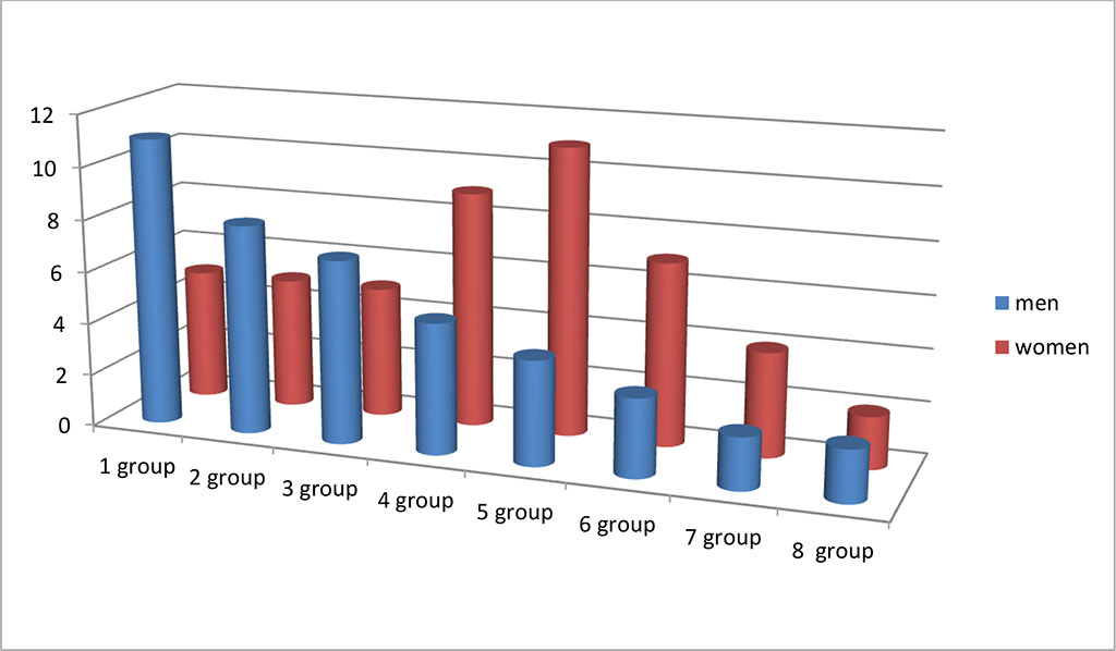
Figure 1 - the frequency of injuries in men and women in terms of age
Based on prospective, controlled comparative studies, monitoring of reparative skin regeneration in patients of various age groups was carried out in accordance with the principles of evidence-based medicine. Classical clinical and morphological research methods were used to monitor reparative processes in damaged skin with subsequent analysis of the data obtained. The study was conducted with the permission of the Ethics Committee of the FSAEI HE FEFU.
Results of own researches. The analysis of the clinical material showed that in the structure of complications after injuries, the violation of skin regeneration in women aged 45 to 65 years prevailed. According to the features of localization of skin lesions in men and women, there are differences in the topography of lesions. Burns and injuries are more present in the group of male patients, in women there were complaints about the complications of surgical correction and anti-aging invasive treatment, as well as invasive cosmetic interventions and piercing (Figure 2).
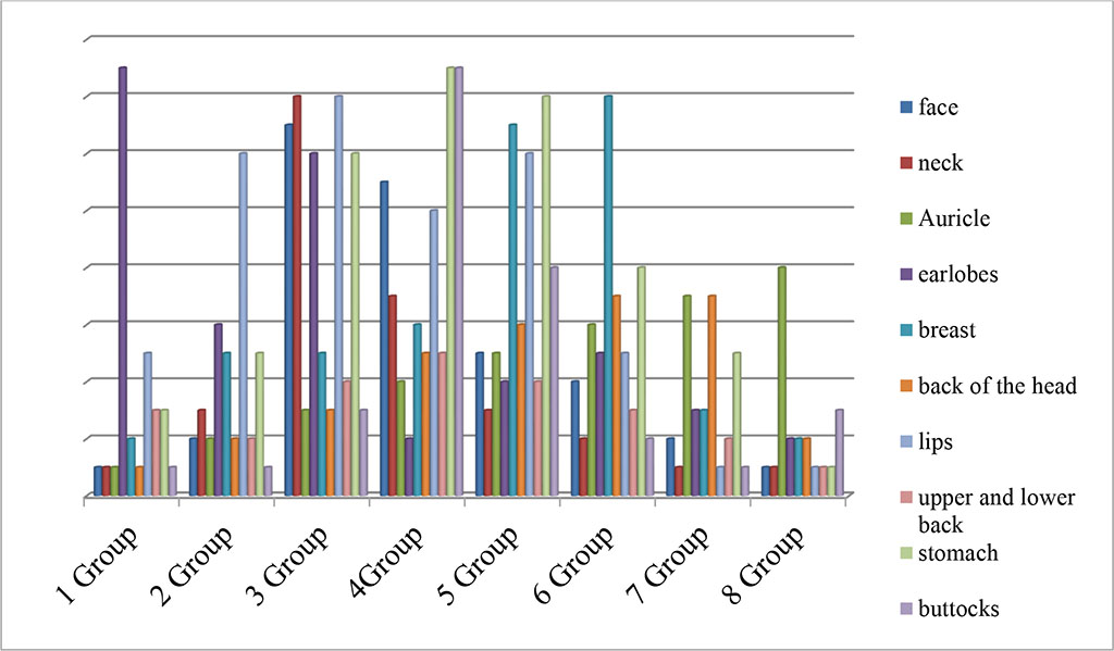
Figure 2 - the frequency of damage to the anatomical zones of the skin in the age aspect
We noted that the most common cause of complications after invasive cosmetic surgery was the use of low-quality implants (Figure 3).

Figure 3 - A) scarring after correction in the zone of the red border of the lips. B) Implants extracted from the lips. C) Keloid scars after otoplasty.
Age-related changes and more frequent complications are due to the fact that in young patients the epidermis has a greater thickness, better trophic supply (more functioning vessels), more collagen, more sebaceous glands, and better expressed skin barrier properties provided by immunocytes and macrophages. With aging, there is a decrease in functioning vessels, an increase in the number of melanocytes, apoptosis is increased, proliferation decreases, and the epidermis becomes thinner.
We noted that the presence of keloid scarring in the anamnesis of patients is an unfavorable factor for the development of pathological scarring. It should be noted that, in general, the healing of damaged skin goes through four specific stages: hemostasis (stopping bleeding), inflammation, proliferation and restitution (overgrowth of the defect by cells due to their creeping onto the defect - restitution), maturation. The complete healing process can take a year or more, depending on the depth and severity of the damage, as well as the age of the patients.
The results obtained contribute to a better understanding of the etiology of skin fibrosis and provide potential etiological target factors for the prevention of fibrotic diseases.

|
||