- Home
- About the Journal
- Peer Review
- Editorial Board
- For Authors
- Reviewer Recognition
- Archive
- Contact
- Impressum
- EWG e.V.
Cite as: Archiv EuroMedica. 2024. 14; 5. DOI 10.35630/2024/14/5.509
Aims: Infectious mononucleosis, caused by the Epstein-Barr virus (EBV), typically presents with fever, exudative pharyngitis and lymphadenopathy. However, atypical presentations can complicate diagnosis and treatment. The objective of this paper is to illustrate the atypical progression of infectious mononucleosis and to delineate the differential diagnosis that should be considered. Additionally, it outlines the therapeutic approaches for managing symptoms associated with the disease.
Case Description: A 25-year-old woman presented at a GP's office with eyelid edema, fever, and significant weight loss. Initial tests and treatments were inconclusive, leading to further investigations. Despite typical treatment for viral infections, the patient’s condition worsened, displaying dyspnoea, acute hepatitis, rash, significantly elevated ESR, hemolytic anemia, leukopenia, and thrombocytopenia. Hospitalization was required for comprehensive care, including steroid therapy and fluid management.
Conclusions: This case highlights the diagnostic challenges posed by atypical presentations of infectious mononucleosis and underscores the importance of considering EBV infection in the differential diagnosis. Early recognition and appropriate management are crucial for optimal patient outcomes.
Keywords: mononucleosis; glandular fever; periorbital edema; hepatitis; differentiation; steroid therapy
Infectious mononucleosis (IM), also known as glandular fever, is caused by primary infection with Epstein Barr virus (EBV), a member of the family Herpesviridae [1]. An alternative name is "kissing disease", as it is transmitted mainly through saliva. Other routes of transmission are sexual intercourse, blood transfusions or organ or bone marrow transplantation. The incubation period for IM is 3 to 6 weeks [2].
Mononucleosis is asymptomatic in many cases; antibodies to EBV are present in about 90% of adults in the general population [3]. In young adults, however, the symptomatic course is relatively more common; a study by Dunmire et al [3] found that among those between the ages of 18 and 22, 75% have typical symptoms, 15% go through the infection atypically, while 10% have no symptoms at all. The data suggest that symptomatic infection in children before puberty is much less common, presumably due to the immaturity of the immune system [3].
When symptoms occur, at least 2 of the 3 are most common: fever, exudative pharyngitis, and lymphadenopathy [4]. The typical course of the disease has two main forms: (1) a sudden onset of symptoms, including severe, intractable sore throat, swollen neck (enlarged lymph nodes) and (2) a gradually increasing feeling of weakness and muscle pain. Most symptoms last up to a maximum of 10 days, but weakness and lymphadenopathy on average remain for about 3 weeks [3].
Approximately 20% of symptomatic patients experience atypical complaints such as abdominal pain, liver and spleen enlargement, nausea, vomiting, swelling of the eyelids and orbital area, and rash [3]. Although subclinical hepatitis manifested by elevated liver enzymes occurs in about 75% of patients, 5-10% develop serious acute viral hepatitis, manifested not only by significant increases in liver enzymes, but also by hepatomegaly or jaundice. In addition, laboratory tests can show leukocytosis and lymphocytosis, with atypical lymphocytes present in the smear [3]. Atypical IM cases are most often seen in specific populations:
Mononucleosis sometimes presents diagnostic challenges for the physician, as it is not difficult to confuse it with strep throat. In case of confusion, when a penicillin derivative is included in the treatment, a typical symptom is the development of a rash all over the body [7].
The disease can result in serious complications, but they occur in less than 1% of cases [8]. The most serious one is a splenic rupture. Others include myocarditis, pancreatitis, pericarditis, airway obstruction, pneumonia, thrombocytopenia, hemolytic anemia, meningitis. Additionally, the so-called "chronic fatigue syndrome" may persist for several months [9]. EBV-associated diseases also include Burkitt's lymphoma and Hodgkin's lymphoma [10], which can be distant complications of infection.
The objective of this paper is to illustrate the atypical progression of infectious mononucleosis and to delineate the differential diagnosis that should be considered. Additionally, it outlines the therapeutic approaches for managing symptoms associated with the disease. Although hospitalization is not necessary in most cases, doctors should be extra vigilant and know when to refer a patient to the hospital.
A 25-year-old woman with chronic gastritis and nontoxic multinodular goiter of the thyroid gland, 1 year after a traffic accident (status post craniofacial surgery; titanium plates in the mandible present) presented to her GP. She gave a history of swelling of the eyelids and tissues around the eyes for 5 days, worsening over time. She had been taking loratadine, with no apparent improvement. She had been running a fever of up to 39.5 °C for 2 days. In addition, she reported increased sweating and a feeling of weakness for about 1 month, during which she had unintentionally lost about 8 kg.
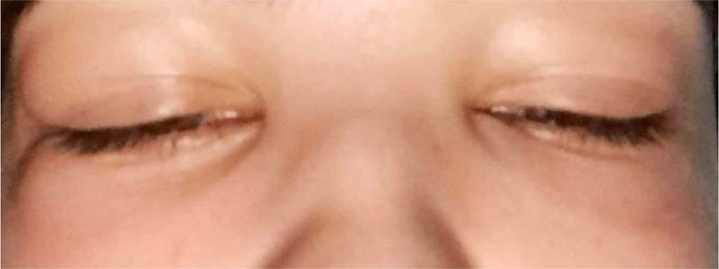
Physical examination revealed a slightly inflamed throat, enlarged palatine tonsils without plaques, swollen nasal cavity mucosa, swollen eyelids and orbital area (Figure 1) and enlarged, non-painful submandibular lymph nodes. Clear to auscultation bilaterally, regular rate and rhythm of the heart, without murmurs nor rubs. Abdomen was soft, non-tender, and not distended. Skin was very moist.

Figure 1. Periorbital edema of the patient.
In the outpatient clinic, a rapid cassette test for influenza, COVID-19, RSV, Mycoplasma pneumoniae, adenovirus was additionally performed, which was negative. Suspecting a possibility of an acute myeloid leukemia or lymphoma, the following tests were ordered urgently: blood count with smear, CRP, creatinine and ALAT. In addition, antipyretics and continuation of loratadine, rest, warm fluids were recommended.
Initial laboratory results showed mild leukopenia and lymphopenia, otherwise no significant abnormalities [Table 1]. A diagnosis of viral infection was tentatively made and a wait-and-see attitude was adopted.
Table 1. The patient's laboratory results on the day of her initial presentation to the GP.
| Examination | Results | Deviation |
| WBC | 3,79 x 10³ /l [4,16 - 11,48] | L |
| RBC | 4,50 x 10⁶/l [3,65 - 5,07] | N |
| HGB | 14.1 g/dl [12.0 - 15.0]. | N |
| HCT | 40,3 % [36,0 - 46,0] | N |
| MCV | 89,6 fl [80.0 - 95.4]. | N |
| PLT | 179 x 103/l [162 - 379] | N |
| # NEU | 2,27 x 10³/l [1,90 - 8,00] | N |
| # LYM | 1,05 x 10³/l [1,20 - 3,30] | L |
| # MON | 0,43 x 10³/l [0,34 - 0,94] | N |
| # EOS | 0,00 x 10³/l [0,04 - 0,41] | L |
| # BAS | 0,03 x 10³/l [0,0 - 1,0] | N |
| # IG | 0,01 x 10³/l [0,1 - 1,0] | N |
| # NRBC | 0,00 x 10³/l [0,0 - 0,1] | N |
| Creatinine | 0,75 mg/dl [0.50 - 0.90] | N |
| GFR | 102 ml/min/1.73m2 [> 60]. | N |
| ALAT | 19 U/I [< 31]. | N |
| CRP | 2,62 mg/l [< 6.00]. | N |
| Peripheral blood smear | Normal peripheral blood picture: RBC, WBC, PLT |
3 days later, the patient again reported to the same GP, citing lack of any improvement, fever up to 40.5 °C increasingly unresponsive to antipyretics, discomfort in the throat, difficulty swallowing, and shortness of breath.
Physical examination revealed increased facial swelling since the last visit, inflamed throat, palatine tonsils even more enlarged still without plaques, enlarged nuchal, occipital, submandibular (massive), cervical, subclavian, inguinal lymph nodes - some palpably tender. Spleen and liver were enlarged for 2 fingers below the rib arch. A fine-spotted diffuse rash was present on the skin of the chest and abdomen.
Repeat laboratory tests were performed [Table 2], which showed a significant worsening - anemia, thrombocytopenia, markedly elevated levels of ALAT and ESR.
Table 2. Results of the patient's laboratory tests after 3 days.
| Examination | Results | Deviation |
| WBC | 3,62 x 10³/l [4,16 - 11,48] | L |
| RBC | 2,34 x 10⁶/l [3,65 - 5,07] | L |
| HGB | 10.8 g/dl [12.0 - 15.0]. | L |
| HCT | 24,5 % [36,0 - 46,0] | L |
| MCV | 105.7 fl [80.0 - 95.4]. | H |
| PLT | 121 x 103/l [162 - 379] | L |
| # NEU | 1,48 x 10³/l [1,90 - 8,00] | L |
| # LYM | 1,84 x 10³/l [1,20 - 3,30] | N |
| # MON | 0,24 x 10³/l [0,34 - 0,94] | L |
| # EOS | 0,00 x 10³/l [0,04 - 0,41] | L |
| # BAS | 0,03 x 10³/l [0,0 - 1,0] | N |
| # IG | 0,01 x 10³/l [0,1 - 1,0] | N |
| # NRBC | 0,00 x 10³/l [0,0 - 0,1] | N |
| Creatinine | 0.71 mg/dl [0.50 - 0.90]. | N |
| GFR | 106 ml/min/1.73 m2 [> 60]. | N |
| ALAT | 195 U/I [< 31]. | H |
| CRP | 22 mg/l [< 6.00]. | H |
| ESR | 61 mm [<22]. | H |
| Peripheral blood smear | Reactive lymphocytes present in lymphocyte population, few visible cloverleaf nuclei. RBCs and PLTs normal |
The patient was referred to the infectious disease ward at a hospital, where a positive serologic test for IgM class antibodies to EBV infection was obtained. The patient denied having any new sexual partners in recent months.
During hospitalization, an abdominal ultrasound was performed, which showed an enlarged liver ( in the midclavicular line about 173 mm), an enlarged spleen measuring 160x40 mm, a dilated uterine body, and free fluid in the cavity of Douglas present.
Additional biochemical tests performed at the hospital showed elevated liver parameters (ALAT maximum of 433 U/L [0-55], ASPAT 360 U/L [5-34], ALP 252 U/L [40-150], GGTP 333 U/L [9-36], lymphomonocytic blood smear, negative tests for HBV, HCV, HIV, electrolyte deficiency.
During the stay, the patient reported very severe fatigue and abdominal distress, lack of appetite, sleep problems, in addition, a persistently elevated body temperature was observed.
Treatment included fluid therapy and electrolytes (Natrium Chloratum 0.9%, Optilyte + Magnesium sulfate, Glucosum 5% i.v.), analgesics and antipyretics (Metamizole i.v.), steroid therapy (Dexaven i.v.), drotaverin, pantoprazole, thiazolidinedioic acid and hydroxyzine.
After a week of hospitalization, the patient was discharged home in a state of optimal improvement (reduction in edema, resolution of fever, decrease in liver enzymes and CRP) with recommendations to lead a quiet, regulated lifestyle, avoid heavy exercise, adherence to an easily digestible diet with total exclusion of alcohol, follow-up laboratory tests at the primary care center in 2 weeks, follow-up ultrasound in 1 month, gynecologic follow-up as soon as possible, and taking dry extract of spotted thistle husk (Silybi mariani fructus extractum siccum).
This case demonstrates what a difficult diagnostic challenge infectious mononucleosis can be. Although usually the first symptom of the disease is a severe sore throat and fever, in the case of the patient in question, the complaints began with a relatively rare periorbital edema. Hence, an antihistamine (loratadine) was included in the treatment, as swelling is relatively often caused by allergies. The fact that the patient had suffered a craniofacial trauma in the previous year resulting in the implantation of a foreign body theoretically might have initially led to ideas that there might be some connection between the two. However, the trajectory of thinking should have changed when the symptoms were joined by a high fever. The patient presented all of the so-called B symptoms: night sweats, fever and unintentional weight loss - symptoms typical of lymphoma [11]. Given that lymphoma can also cause swelling of facial tissues [12], its exclusion was a must for the doctor, hence the order for an urgent blood count with smear. Leukopenia with lymphopenia and a normal blood smear ruled out lymphoma with high probability, directing suspicion toward some kind of viral infection, which the cassette tests performed at the first visit did not show. It is worth noting that mononucleosis typically presents inversely (with leukocytosis and lymphocytosis), so at this stage of diagnosis it would have been difficult to suspect it, especially since the patient did not show severe sore throat, and no effusion was present on the throat, which is relatively common in mononucleosis. A wait-and-see attitude was adopted, ordering symptomatic treatment and expecting that the infection would reduce on its own and the symptoms would begin to withdraw. Since within 3 days the symptoms not only did not reduce, but there was a significant worsening, the fever stopped responding to medications, the massive palatine tonsils significantly restricted the ability to breathe freely causing shortness of breath and laboratory tests significantly worsened (ESR = 61 mm, thrombocytopenia, a sharp drop in Hgb) hospitalization was necessary. In the hospital, the differential diagnosis was deepened. Leukopenia with lymphopenia, generalized lymphadenopathy, sore throat, fever, splenic enlargement, and a rash without prior exposure to amoxicillin required exclusion of mononucleosis-like syndrome in the course of HIV [13]; hence the inclusion of questions about sexual behavior in the history and also the ordering of HIV testing. Because of the hepatomegaly, it was also worthwhile to rule out acute hepatitis B and C. Finally, serological testing of IgM class antibodies for EBV infection complicated acute hepatitis (more than 10-fold elevation of liver enzymes ALAT and ASPAT, significantly elevated GGTP and ALP). The above case showed how such severe cases of the disease are treated - although normally mononucleosis does not require steroid therapy, in the case of airway obstruction by swollen tonsils, steroids can be very helpful. The patient should also be protected from dehydration, hence fluid therapy is often necessary. For abdominal pain, decongestants such as drotaverin are useful, while thiazolidinedioicarboxylic acid can be used to help with liver damage. Once the disease is under control, it is crucial to advise the patient to rest and avoid exercise - as life-threatening splenic rupture with subsequent internal hemorrhage can occur if these recommendations are not followed.
In conclusion, infectious mononucleosis in some cases can be very difficult to diagnose, and should be differentiated from allergic reactions, bacterial tonsillitis, influenza, RSV infection, COVID-19, Mycoplasma pneumoniae, adenovirus, lymphoma, HIV infection or HBV and HCV, among others. A useful diagnostic tool is serologic antibody testing in the IgM class for EBV infection. In cases of fever unresponsive to antipyretic and anti-inflammatory therapy, respiratory obstruction or overt acute hepatitis, hospitalization may be necessary.