- Home
- About the Journal
- Peer Review
- Editorial Board
- For Authors
- Reviewer Recognition
- Archive
- Contact
- Impressum
- EWG e.V.
Cite as: Archiv EuroMedica. 2024. 14; 5. DOI 10.35630/2024/14/5.508
The aim: To improve the effectiveness of diagnosing and developing treatment strategies for patients with the vestibular position of permanent canines
Material and Methods: Material for the conducted research were the database obtained during clinical examination of 7172 patients that have been referring to our clinic for the orthodontic treatment.
Of these, 2,669 were male (37.21%) and 4,503 were female (62.79%). Among the total number of examined patients, 461 were found to have a vestibular position of tooth 13 (6.43±0.29%), and 357 patients had a vestibular position of tooth 23 (4.98±0.26%). A combination of vestibular positions of teeth 12 and 13 was found in 553 patients (7.71±0.31%).
Simulation of various orthodontic treatment options to achieve the optimal aesthetic result using the "Dolphin Imaging 11.9" program was conducted in two scenarios: one with the removal of the first permanent premolars and the other without tooth extractions, using maxillary expansion and distalization of the posterior teeth. Two clinical cases are illustrated to demonstrate the stages of treatment in patients with vestibular positioning of maxillary canines, both with and without tooth extraction.
Results: The author concludes that the use of the "Dolphin Imaging 11.9" program significantly enhances the effectiveness of diagnosing and orthodontically treating patients with vestibular positioning of permanent maxillary canines, helping to achieve harmony between skeletal and soft tissue facial relationships.
Keywords: vestibular positioned canines, Dolphin-Imaging, soft tissues facial profile.
The vestibular (ectopic) displacement of maxillary permanent canines is one of the most common conditions observed in patients requiring orthodontic treatment. The primary concern of these patients is the problems of facial and smile aesthetics. According to literature, the frequency of vestibular positioned canines has a 2 - 3,06% average in general prevalence of dentofacial anomalies [1-5].
The macro dens of incisors, crowding of teeth, mesial shift of posterior group of teeth, decrease of the transverse and sagittal sizes of jaws, etc. and can be the etiological factors promoting emergence of the ectopic eruption of maxillary permanent canines [6-8].
So far there is no consensus concerning a choice of tactics of treatment of patients with this pathology. Some authors in case of vestibular positioned of permanent canines as a variant of treatment recommend extracting the first premolars, others consider it expedient to carry out treatment by expansion of jaws and a distalisation of posterior teeth [9 - 11]. Such variety of opinions concerning the same question depends, first of all, on exact diagnostics of a condition of all parameters of dentofacial system and the subsequent right choice of the corresponding plan of orthodontic treatment.
Research objective – improvement of diagnostics and tactics of orthodontic treatment in patients with the vestibular positioned maxillary permanent canines.
Material for the conducted research were the database obtained during clinical examination of 7172 patients that have been referring to our clinic for the orthodontic treatment.
The detailed examination of patients gave us the opportunity to create a robust database on the prevalence of dentofacial anomalies, including the occurrence of vestibular positioned maxillary permanent canines. Out of the total number of surveyed patients, 2,669 (37.21%) were males, and 4,503 (62.79%) were females. The ectopic positioning of the tooth 13 was found at 461 patients that is 6,43±0,29%, tooth 23 - at 357 (4,98±0,26%), the simultaneous vestibular eruption of both maxillary canines at 553 people (7,71±0,31%).
During examination of all patients clinical, radiological, biometric, statistical methods of research were applied.
For the statistical analysis selective shares (p) and their mistakes (mp) were calculated at alternative group option. For comparison of the received results Χ2 criterion (Pearson consent criterion) was used.
Prevalence of vestibular eruption of permanent canines at patients depending on ratios of their dentitions was also found. At a sagittal ratio of dentitions and class I, the ectopic eruption of canines is found at 3545 patients - (49,43±0,59%), a class II – at 2710 patients - (37,79±0,57%), a class III at 917 patients - (12,79+0,39%). At vertical ratio same anomaly of canines was found at 891 patients - (12,42±0,39%) with an open bite, at 1629 patients - (22,71±0,49%) with a deep bite.
The highest incidence of abnormal eruption of canines was observed in Class I patients (49.43%), indicating a higher likelihood of this problem with normal occlusal relationship.
In a contemporary orthodontics for diagnostics and treatment planning the new software is applied, allowing not only to choose optimum methods of diagnostics and treatment, but also to predict results regarding further growth and development of structures of dentofacial system.
For accurate diagnostics and optimal treatment planning, we used the software Dolphin Imaging 11.9, which offers a key advantage by creating an individualized orthodontic treatment plan based on a specialized algorithm. This plan takes into account the patient's skeletal jaw relationships and soft tissue profile, ensuring personalized treatment for each patient. Simulation of various options of orthodontic treatment and receiving optimum esthetic result by means of the “Dolphin-Imaging-11,9” software was carried out in two options according to the scheme:
In cases with convex profile and protrusive lip position beyond Ricketts' E-line, if simulation using Dolphin software indicated that extraction would improve the profile, we recommended the option of extracting first premolars.
In cases of concave profile if extraction of the first maxillary premolars worsened a situation or maxillary expansion improvement became acceptable, non-extraction treatment is recommended for the patients.
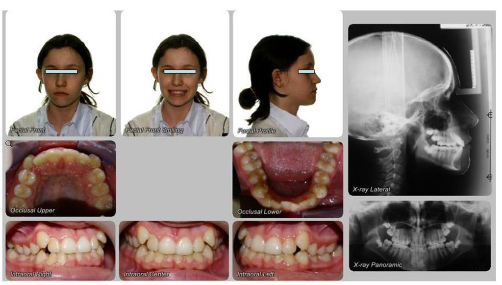
Patient L.V., aged 11, Case No. 3087, presented with concerns regarding the vestibular positioning of the maxillary canines. Clinical examination revealed Class I malocclusion, mandibular crowding, and vestibular displacement of the maxillary canines. A panoramic radiograph confirmed insufficient space for the maxillary permanent canines and the presence of developing third molar buds (Fig. 1).

Fig. 1 Clinical and radiographic view of 11-year-old girl’ dentition before treatment.
Before treatment the patient had convex profile (Fig. 2a). At various options of computer simulation – with extraction and non-extraction it is visually seen that if we plan treatment with expansion of jaws a profile worsens (Fig. 2b), and in a case of extraction of the first premolars the patient’s profile considerably improves (Fig. 2c).

Fig 2 Comparison of profile photos while making treatment forecast. a - before treatment; b – treatment forecast with expansion of jaws – profile deterioration; c - treatment prediction with extraction of four premolars - profile improvement.

Fig. 3 Stages of orthodontic treatment.
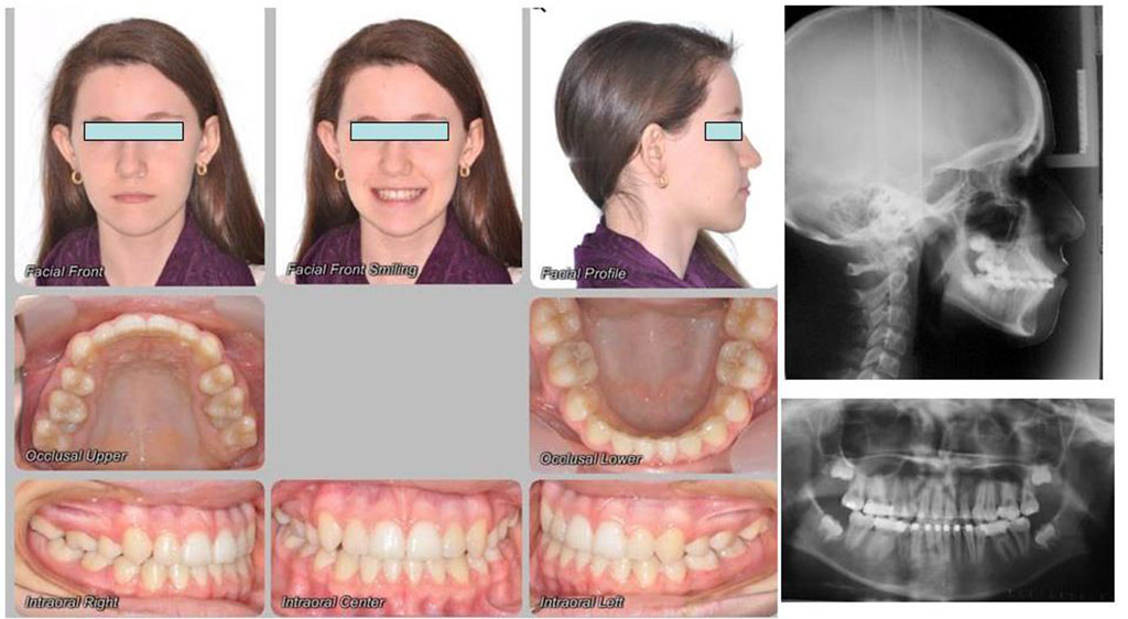
Treatment is completed with the establishment of proper fissure-to-cusp contacts (Fig.4).

Fig. 4 Clinical and radiographic view of a 13-year-old female patient's post-treatment dentition.
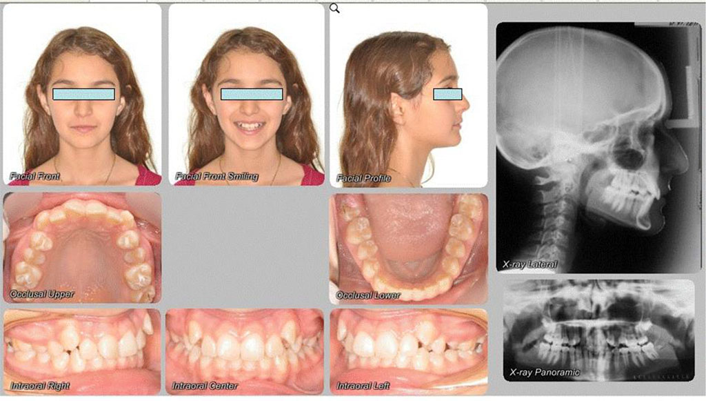
Patient R.A., aged 13, Case No. 4836, presented with complaints regarding an aesthetic defect. Clinical examination revealed an Angle Class II malocclusion, maxillary anterior crowding, and ectopic eruption of the maxillary permanent canines. A panoramic radiograph indicated insufficient space for the maxillary canines and the presence of developing third molar germs (Fig. 5).

Fig.5 Clinical and radiographic view of 13-year-old girl's dentition before treatment.
Before treatment the patient had concave profile, lips were at backward position to the esthetic line that is presented in Fig. 6a. At various options of computer simulation – with extraction and non-extraction it is notable that if we do extraction, a profile (fig. 6b) worsens, in a case with expansion of jaws the patients’ profile considerably improves (Fig. 6c)

Fig. 6 Comparison of 13-year-old female patient’s photos during treatment outcomes prediction. A – before treatment; b – treatment simulation with extractions indicates potential profile deterioration; c – treatment simulation with jaw expansion demonstrates potential profile improvement
The stages of orthodontic treatment using self-ligating system “Quick” – leveling of dental arches, the use of class II elastics for creation of interproximal contacts (fig. 7a-c).

Fig.7 Progression of leveling and formation of multiple occlusal contacts.
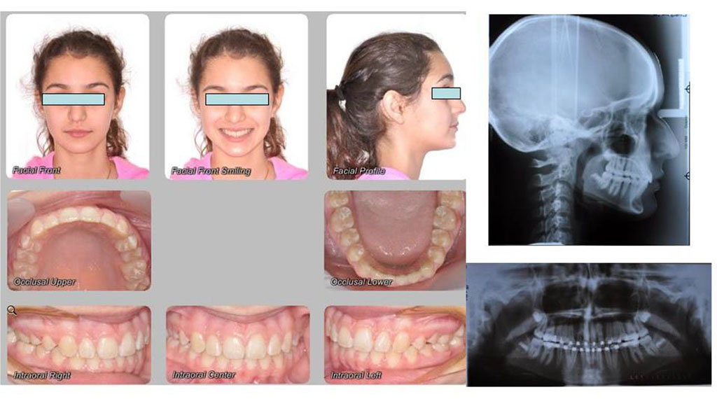
The treatment is completed with the achievement of maximum intercuspation, ideal fissure-tubercle contacts are reached (fig. 8). The achieved profile corresponds to the planned outcome.

Fig.8 Clinical and radiographic view of 1year post-treatment dentition.
Digital diagnostics, utilizing three-dimensional imaging of dental arches, bone structures, and the face, enables comprehensive treatment planning in three dimensions, starting from the most superficial layers. This approach ensures greater precision in identifying anatomical details, enhancing the accuracy and effectiveness of orthodontic interventions [11, 12]. Computer-based diagnostics, treatment planning, and outcome prediction in orthodontics have fundamentally transformed approaches to care and achieving optimal aesthetic results. The traditional analysis of X-rays, clinical data, and diagnostic models is no longer sufficient for accurate diagnosis and treatment selection. Modern computational algorithms process cephalometric radiographs and 3D images, allowing for the identification of balanced facial profiles [13]. This enables more detailed and data-driven treatment planning, providing personalized and more effective orthodontic solutions for each patient.
Thus, it is possible to note that application of the rational software "Dolphin-Imaging 11,9" sub substantially improves effectiveness of diagnostics and orthodontic treatment in patients with the ectopic displacement of maxillary permanent canines and allows to achieve harmony in ratios between skeletal and facial soft tissues.