- Home
- About the Journal
- Peer Review
- Editorial Board
- For Authors
- Reviewer Recognition
- Archive
- Contact
- Impressum
- EWG e.V.
The aim of our study was to investigate the cellular composition of the lymphoid structures of the human intrahepatic bile ducts. Materials and Methods: The object of the study was the lymphoid structures of the walls of the intrahepatic bile ducts obtained from 45 cadavers of different ages. Lymphoid nodules in the walls of the intrahepatic bile ducts were investigated by T. Hellman’s method. The microanatomy of lymphoid structures was studied on microscopic preparations. Microscopic preparations were stained according to Van Gieson, with hematoxylin-eosin, methylene blue. The digital data obtained during the study was subjected to statistical processing. Results: A macromicroscopic study of lymphoid formations in the intrahepatic bile ducts showed that lymphoid formations in the walls of these organs are represented by lymphoid nodules and diffuse lymphoid tissue. The peripheral contours of the lymphoid nodules are clearly defined. Histologically, the cellular composition of lymphoid nodules and diffuse lymphoid tissue was studied. In the mucous membrane and in the submucosa, small, medium sized lymphocytes, and large lymphocytes, macrophages, plasma, reticular, mast cells, cells in a stage of mitosis, degeneratively altered (destructive), and other forms of lymphoid cells are determined. Conclusions: According to our data, newborns have not only lymphoid nodules, but also a typical cellular composition in the lymphoid tissue, which has a completely “mature character”. The results of morphometric studies showed that in early childhood there is a high percentage of small, medium, large lymphocytes, macrophages, cells with signs of mitosis.
Keywords: intrahepatic bile ducts; lymphoid nodules; diffuse lymphoid tissue; lymphocytes; mast cells.
Diseases of the gallbladder and biliary tract are considered an important medical and social problem. Recent studies show that the number of various pathologies in this area has been constantly increasing. The nomenclature of diseases of the digestive system was replenished with "new" diseases, such as cholesterolosis of the gallbladder, anomalies of the biliary tract, etc. [6,10,15].
The successful development of biliary surgery has always been a stimulus for a detailed topographic and anatomical study of the hepatopancreatoduodenal zone, with the majority of works traditionally devoted to the anatomy of the extrahepatic bile ducts [5,11]. However, information on the morphology of the intrahepatic bile ducts is currently insufficient [9].
Regardless of organ affiliation, lymphoid formations are considered the source of many diseases [4]. Lymphoid structures of various organs have not been studied widely enough. The lymphoid structures of the intrahepatic bile ducts have been especially poorly studied.
The aim of our study was to study the cellular composition of the lymphoid structures of the human intrahepatic bile ducts.
The object of the study was the lymphoid structures of the walls of the intrahepatic bile ducts obtained from 45 cadavers of different ages at death. Lymphoid nodules in the walls of the intrahepatic bile ducts were studied by the method developed by Hellmann T. [7]. The microanatomy of lymphoid structures was studied on microscopic preparations. Microscopic preparations were stained according to Van Gieson, hematoxylin-eosin, methylene blue [14]. The digital data obtained during the study was subjected to statistical processing. In this study, we adhered to ethical principles of biomedical research. Statistical analysis was carried out in MS Excel 2016 and IBM Statistics SPSS 26 programs [3].
A macromicroscopic study of lymphoid formations in the intrahepatic bile ducts shows that lymphoid formations in the walls of these organs are represented by lymphoid nodules and diffuse lymphoid tissue. According to our data, lymphoid nodules on total preparations of the intrahepatic bile ducts are detected as dark (mainly dark blue) structures located against a lighter background of the surrounding wall of the organ. The peripheral contours of the lymphoid nodules are clearly defined (Fig.).
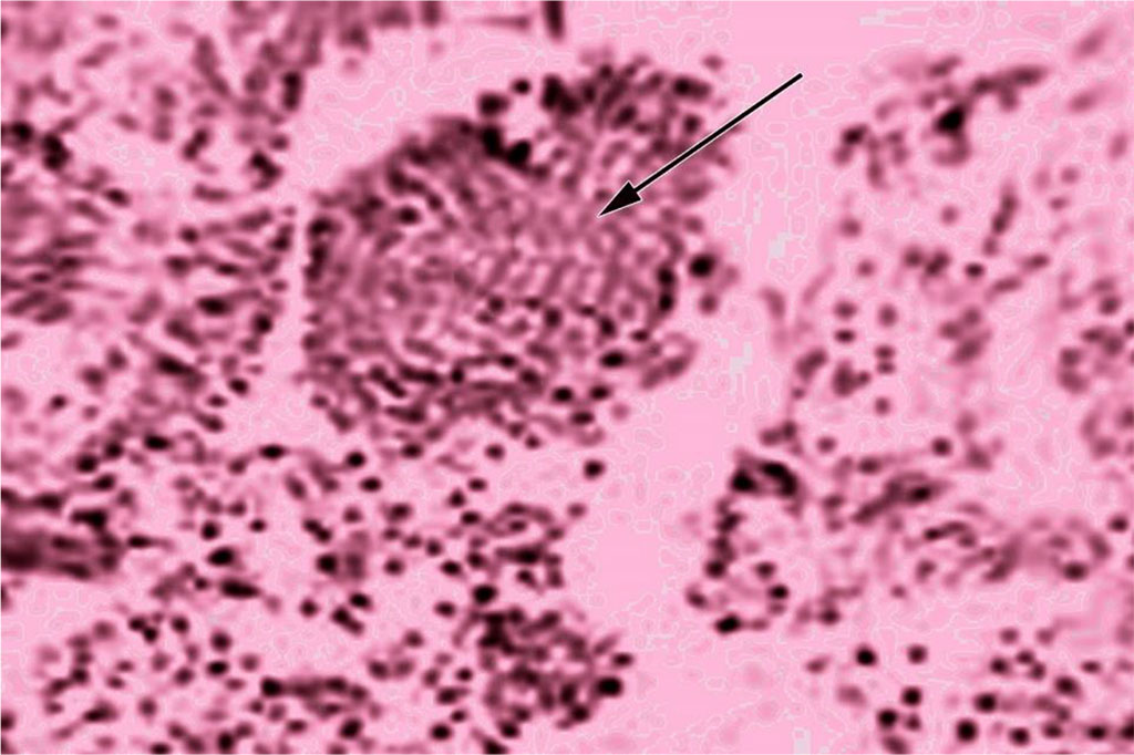
Fig. Lymphoid nodules in the intrahepatic bile duct, transverse section. Hematoxylin-eosin staining. Magnification 190x
Histologically, we studied the cellular composition of lymphoid nodules and diffuse lymphoid tissue. In the mucosa and submucosa, small, medium, and large lymphocytes, macrophages, plasma, reticular, mast cells, cells in a state of mitosis, degeneratively altered (destructive), and other forms of lymphoid cells are determined.
Neutrophils are occasionally detected
in lymphoid tissue.
On
histological sections, we studied the percentage of different types
of lymphoid cells that form diffuse lymphoid tissue in the walls of
these organs (Table).
Table: Cellular composition of lymphoid diffuse tissue in the walls of the trachea and main bronchi in samples of different ages
| Сell type | Age, number of cells (in%) | ||||
| Newborns | Early childhood | Puberty | Adulthood | Old age | |
| Small lymphocytes | 64,20,65 (62-68) | 64,60,71 (62-68) | 60,42,12 (52-68) | 60,04,41 (52-68) | 56,31,2 (50-62) |
| Medium sized lymphocytes | 15,20,65 (12-17) | 13,20,59 (11-16) | 13,20,64 (11-17) | 13,60,70 (11-18) | 13,60,97 (10-19) |
| Large lymphocytes | 2,40,30 (1-4) | 3,50,30 (1-4) | 4,00,30 (2-5) | 3,50,47 (1-6) | 3,00,47 (1-6) |
| Reticular cells | 10,20,30 (8-11) | 8,10,30 (6-9) | 10,20,43 (8-12) | 10,20,47 (8-13) | 10,40,65 (8-14) |
| Plasma cells | 1,20,30 (0-3) | 1,10,22 (0-2) | 1,20,22 (0-2) | 1,20,22 (0-2) | 1,20,22 (0-1) |
| Macrophages | 1,00,22 (0-2) | 1,00,22 (0-2) | 2,50,22 (1-3) | 2,80,30 (1-3) | 5,20,30 (4-7) |
| Cells in a stage of mitosis | 1,20,22 (0-2) | 2,80,30 (2-5) | 2,00,30 (1-4) | 2,00,22 (1-3) | 1,60,30 (1-4) |
| Degenerative cells | 0,50,13 (0-1) | 0,50,13 (0-1) | 2,40,47 (1-5) | 2,80,30 (1-4) | 4,50,30 (3-6) |
Note
1: 5 cases were taken in each group
Note
2. This table shows the number of reticular cells in diffuse lymphoid
tissue
The percentage of small lymphocytes is maximal in early childhood. It is slightly higher than in newborns: 1.01 times (p> 0.05). In early childhood, it is higher than in puberty (1.07 times, p>0.05), the 1st period of adulthood (1.08 times, p>0.05) and senile age (1.14 times,p>0.05).
The percentage of medium-sized lymphocytes is maximum in newborns (15.2%), in comparison with which in early childhood this indicator decreases by 1.15 times (p> 0.05), does not change in puberty, decreases in the 1st period of adulthood, and decreases in senile age by 1.12 times (p>0.05).
Compared with newborns, the percentage of large lymphocytes in early childhood increases by 1.46 times (p<0.01), in puberty by 1.67 times (p<0.001), in the 1st period of adulthood by 1.45 times (p<0.01), in senile age by 1.25 times (p<0.05).
The percentage of reticular
cells in diffuse lymphoid tissue compared with newborns in early
childhood is reduced by 1.26 times (p<0.05). In puberty and the
1st period of adulthood, the level of these cells corresponds to the
neonatal period and then slightly increases.
The
percentage of plasma cells in diffuse lymphoid tissue is kept at the
same level.
The number of mast cells in comparison with the neonatal period in early childhood increases by 1.09 times (p>0.05). Compared with early childhood, this indicator decreases in puberty by 1.22 times (p<0.05), in the 1st period of adulthood by 1.24 times (p2<0.05), in senile age by 1.25 times (p<0.05).
The percentage of macrophages in the diffuse lymphoid tissue of the trachea and main bronchi compared with newborns and in early childhood in puberty increases by 2.50 times (p<0.001), in the 1st period of adulthood by 2.80 times (p< 0.001), and in senile age by 5.20 times (p<0.001).
Compared with the number of cells in a stage of mitosis in the neonatal period in diffuse lymphoid tissue, their number in early childhood increases by 2.33 times (p<0.001), in puberty and in the 1st period of adulthood by 1.66 times (p< 0.001), and in senile age by 1.33 times (p<0.01).
In comparison with the number of degenerated cells of the lymphoid row in the neonatal period and in early childhood (0.5%), the content of these cells in puberty increases by 4.80 times (p<0.001), in the 1st period of adulthood by 5.60 times (p<0.001), in senile age by 9.0 times (p<0.001).
We have proved that the lymphoid formations present in the walls of the intrahepatic bile ducts are represented by lymphoid nodules and diffuse lymphoid tissue. At the same time, we are not inclined to believe that there are lymphoid accumulations (prenodules) in the walls of these organs. According to our data, all compact lymphoid formations of the intrahepatic bile ducts have clear peripheral contours, which allows them to be classified as typical lymphoid nodules.
According to the data obtained, the cellular composition of the lymphoid structures of the intrahepatic bile ducts corresponds to that of the peripheral organs of the immune system [1,2,8,12,13]. So, in the composition of the lymphoid structures of these organs, small, medium-sized, and large lymphocytes, plasma cells, macrophages, mast cells, and other cells of the lymphoid series are determined.
According to our data, newborns have not only lymphoid nodules but also a typical cellular composition in the lymphoid tissue, which has a completely "mature character".
The results of morphometric studies showed that in early childhood there is a high percentage of small, medium-sized, and large lymphocytes, macrophages, and cells with signs of mitosis, which indicates a high activity of lymphocytopoietic processes.
Thus, lymphoid formations in the walls of the intrahepatic bile ducts are represented by lymphoid nodules and diffuse lymphoid tissue. In the composition of the lymphoid structures of these organs, small, medium-sized, and large lymphocytes, plasma cells, macrophages, mast cells, and other cells of the lymphoid series are determined. The maximum development of lymphoid formations occurs in early childhood.

|
||