- Home
- About the Journal
- Peer Review
- Editorial Board
- For Authors
- Reviewer Recognition
- Archive
- Contact
- Impressum
- EWG e.V.
Cite as: Archiv EuroMedica. 2022. 12; 4: e1. DOI 10.35630/2199-885X/2022/12/4.15
Background. Over the past 20 years, the prevalence of wedge-shaped defects has increased by 14%, and in some countries of the world it reaches critical values - 82%. At the same time, the treatment of this pathology is carried out at the later stages of its development with excision of the hard tissues of the tooth and subsequent restoration, without taking into account the initial macro- and microstructural changes in the enamel. The purpose of our study was an experimental in vitro study of anatomical and morphological features of the tooth enamel structure, its qualitative and quantitative composition in the initial forms of a wedge-shaped defect.
Materials. As the material for our study we used intact teeth removed for orthodontic indications and with initial forms of a wedge-shaped defect of classes 1 and 2 according to the classification of Shevelyuk Yu.V., Makeeva I.M. (2011), subsequently 63 sections were made out of these teeth.
Methods: clinical, experimental, scanning electron microscopy and energy dispersive analysis, statistical, analytical.
Results. Structural transformations of enamel were revealed in the initial forms of the wedge-shaped defect, the boundaries of the lesion were determined, and morphological changes were established in the tissues adjacent to the lesion involved in the pathological process against the background of quantitative and qualitative indicators of its chemical composition.
Conclusion. a comparative analysis of the enamel microstructure in normal and wedge-shaped defects confirms the heterogeneity of the lesion and the transformation of the chemical, quantitative and qualitative composition of the enamel.
Keywords: wedge-shaped defect, enamel microstructure, enamel elemental composition
Wedge-shaped defects of hard dental tissues are quite common among diseases of non-carious origin. Diagnosis and treatment of wedge-shaped defects of teeth are challenging due to their prevalence, controversy of views on the origin and cause of the disease, its tactics of treatment and poorly reporting in the medical literature [5-7, 10-12]. Restoration of tooth tissues with such defects presents certain difficulties, as a special approach to dissection, treatment, and the right choice of filling material is required. Besides, the general diseases of the body, which affect the process of loss of dental hard tissue, should be taken into account [16-18].
In the structure of dental morbidity in Russia, non-carious lesions of hard dental tissues take a significant place supporting the global trend towards further growth and rejuvenation - 82% [1-4].
Diagnosis of a wedge-shaped defect is most often carried out at the later stages of its development, and treatment is carried out without taking into account the pathogenetic mechanisms of its onset and consists in excision of damaged tooth tissues with subsequent restoration [13-15]. Therefore, dental interventions are aggressive and often lead to complications and adverse outcomes [8, 9].
Issues related to characteristics of the pathogenetic mechanisms for the development of wedge-shaped defects are still debatable and require further study.
Aim: To clarify the anatomical and morphological features of the tooth enamel structure, its qualitative and quantitative composition in the initial forms of the wedge-shaped defect in the conditions of the experiment in vitro.
To achieve this goal, 68 sections of intact teeth (5) were studied as well as teeth affected by a wedge-shaped defect (58) within the limits of enamel of class 1 (25 samples) and class 2 (33 samples) (Shevelyuk Yu.V., Makeeva I.M. 2011).
The sections were prepared according to a special technology, by separating the vestibular surface of the crown in the direction from the cutting edge to the neck along the central axis, with constant water and air cooling. The transverse section of the tooth crown was processed with abrasive discs. The resulting biological product was washed in distilled water, placed into the ultrasonic bath for 30 minutes.
With the help of scanning electron microscopy (scanning electron microscope Quanta 200i 3D FEI) at the Laboratory of electron microscopy and small-angle X-ray diffractometry (Department of General Physics, N. P. Ogarev National Research Mordovian State University) the microstructure of enamel in the affected area and tissues adjacent to the lesion were studied. The qualitative and quantitative composition of the enamel of intact teeth and teeth with initial forms of a wedge-shaped defect was studied using the method of energy dispersive microanalysis.
The results obtained underwent statistical processing.
As a result of the study, 128 electronic images of samples of intact and affected teeth with a wedge-shaped defect were obtained and analyzed.
Microphotographs of intact teeth sections obtained using SEM visualize a relatively uniform, even, smooth surface of the enamel layer (Fig. 1).
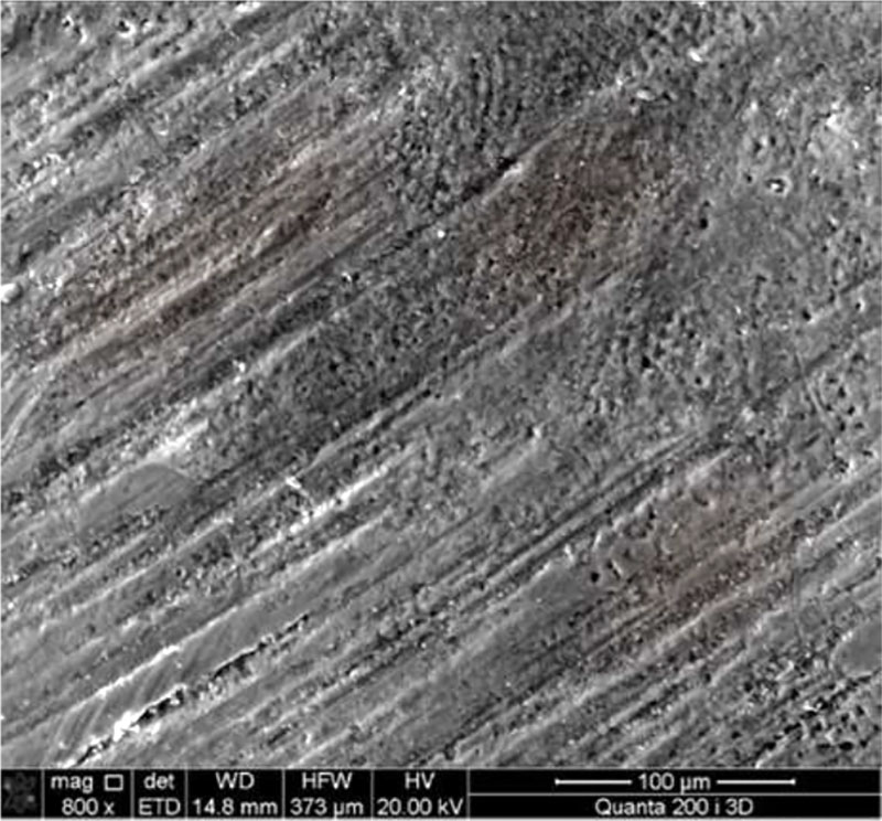
Figure 1 - The enamel surface of an intact tooth, SEM magnification 800
In addition, there is a homogeneous structure of enamel with various types of arrangement of the heads of enamel prisms. There are areas with a sinking and protruding position of the heads of enamel prisms, alternating bands located horizontally at a relatively equal distance from each other. Due to this, the enamel acquires individual characteristics - microroughness, giving it the appearance of transverse striation (Figure 2).
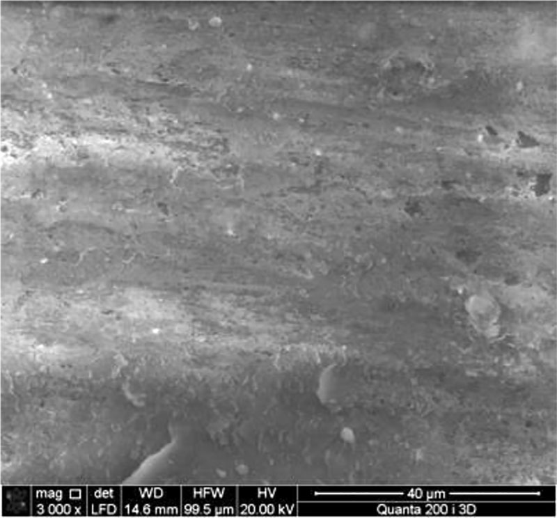
Figure 2 - Microstructure of an intact tooth, enamel prisms, SEM, magnification 3000
Quantitative and qualitative indicators of the microelement composition of intact enamel are represented by the following elements: C - 19.68±1.5%; N- 4.41±0.76%; O - 30.57±1.3%; F - 0.26±0.01%; Na - 3.41±0.4%; K-1.64±0.21%; P - 12.62%±0.9; Cl -2.02%±0.2; Ca - 25.27±1.9% (Fig. 3).
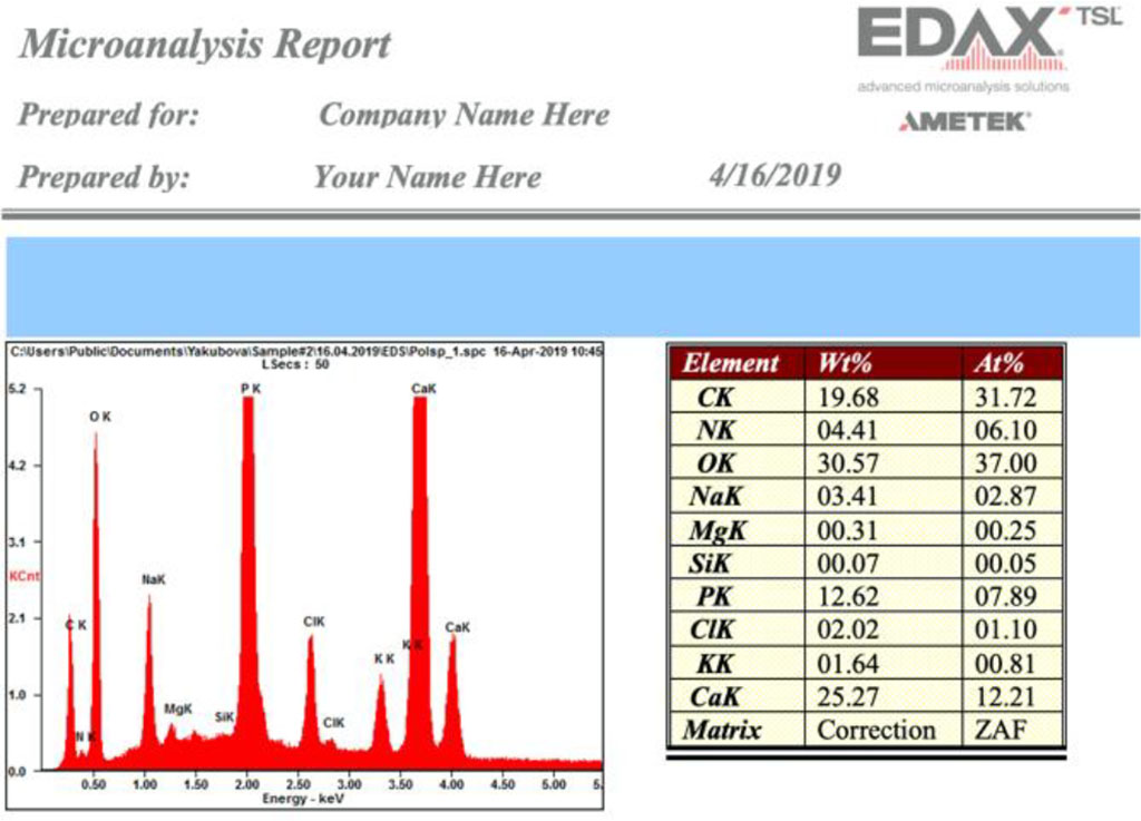
Figure 3 - Elemental composition of intact enamel
The study of electronic images of the pathological focus in wedge-shaped defect with magnification made it possible to identify three different damage zones in the structure: the pathological focus (demineralization zone), the border of the pathological focus and the compromised zone of apparently unchanged enamel (Fig. 4).
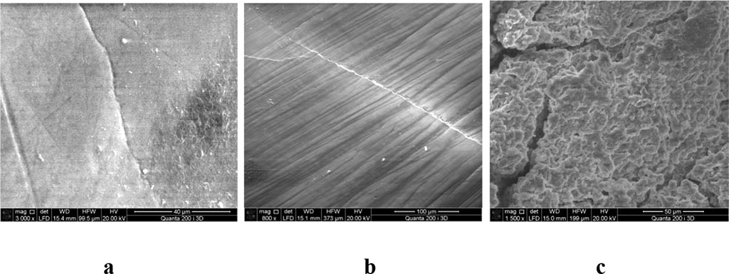
Figure 4: a. The surface of the enamel in the lesion with a wedge-shaped defect, SEM, magnification 3000; b. The surface of the enamel on the border with a wedge-shaped defect, SEM, magnification 800; c. The surface of the intact enamel on the border with the wedge-shaped defect, SEM, magnification 15000
In the first zone on the surface of the enamel in the focus of the wedge-shaped defect, the presence of cracks along the enamel prisms is determined, their width varies, their location and distribution is chaotic. In the focus, pores of different diameters are noted.
Enamel, bordering with a zone of a wedge-shaped defect, was visualized as covered with multiple chaotically located, horizontal, linear defects, cracks, passing along the border of enamel prisms.
The study the enamel layer areas of the tooth hard tissues, damaged by a wedge-shaped defect at the level of intact enamel, located near the border of the defect itself, revealed destructive changes in the form of micropores of different diameters, cracks of different depths and directions, foci of chaotic striation.
In the focus of enamel lesions with a wedge-shaped defect, the chemical elements corresponded to a certain qualitative and quantitative composition: C - 22.27 ± 1.5%; N- 7.92 ± 0.76%; O - 38.22 ± 1.3%; Na - 0.62 ± 0.04%; Р — 10.09 ±0.9%; Cl -0.26±0.06%; Ca - 19.88 ± 1.9%, Mg - 0.52 ± 0.04% (Pic. 5).
In the area adjacent to the focus of the lesion with a wedge-shaped defect, the quantitative and qualitative composition of the microelements of the tooth enamel looked in numerical terms as follows: C - 7.68 ± 2.4%; N- 2.3±0.4%; O — 41.43±1.4%; Na - 0.82% ± 0.02; Р — 12.1±1.5%; Cl -0.44±0.01%; Ca - 29.61 ± 3.9%, K - 0.24 ± 0.13%.
The quantitative and qualitative composition of the chemical elements of the tooth enamel, damaged by a wedge-shaped defect in the intact enamel, corresponded to the following set: C - 27.2 ± 1.4%; O - 32.09 ± 3.4%; Na - 0.79% ± 0.65%; Р — 15.1±1.5%; Cl -0.41±0.25%; Ca - 26.21 ± 3.9%, N - 5.1 ± 0.13%.
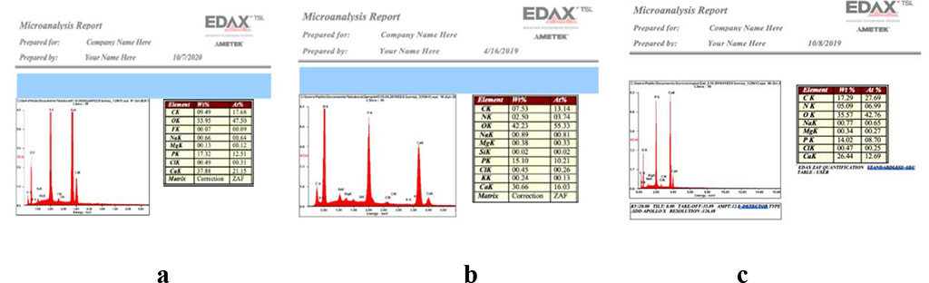
Figure 5 - Study of the chemical composition of enamel a. in the lesion with a wedge-shaped defect; b. the border of the pathological focus; c. The surface of intact enamel on the border with a wedge-shaped defect
A paired two-sample t-test for averages in a comparative analysis of the chemical composition of the intact enamel surface and with an affected wedge-shaped defect showed that the pathological process contributes to an increase in the amount of oxygen by 2.3 times, a decrease in the number of carbon compounds by 1.6 times, against the background of a decrease in the content fluorine by 4.5 times (statistically significant, p <0.05). Microelements such as sulfur appear in the composition - 0.99 ± 0.01, which exceeds its normal content. There is an increase in the nitrogen content by 2.8 times, and the amount of calcium decreases by 1.9 times. The ratio of phosphorus-calcium is 1:0.7, which indicates the process of demineralization (Fig. 6).
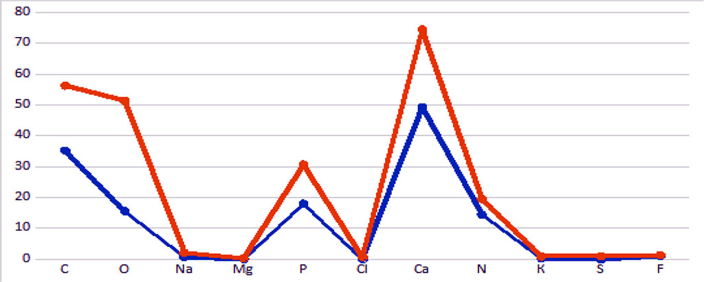
Figure 6. Dynamics of average data for intact and damaged enamel with a wedge-shaped defect
As a result of the study, for the first time, structural changes in tooth enamel were studied in the initial stages of a wedge-shaped defect, the boundaries of the lesion were described, and morphological changes in the tissues adjacent to the lesion involved in the pathological process were identified. It has been established that in class 1 and 2 wedge-shaped defects, microstructural transformations of the enamel are observed not only in the lesion, but also in apparently healthy enamel areas adjacent to it, as well as changes in the qualitative and quantitative elemental composition.
Changes in the qualitative and quantitative indicators of the microelement composition in the zone of the pathological focus are characterized by an increase in the number of oxygen by 2.3 times, a decrease in the amount of carbon compounds by 1.6 times, a decrease in fluorine by 4.5 times, a decrease in calcium by 1.9 times, with the presence of trace elements not typical for healthy enamel, such as sulfur.
The revealed changes in the lesion and in the adjacent tissues indicate a deep enamel lesion in a wedge-shaped defect, which is not taken into account in the current treatment protocol. The data obtained indicate the need to search for and develop broader, combined methods for the treatment of wedge-shaped defects.

|
||