- Home
- About the Journal
- Peer Review
- Editorial Board
- For Authors
- Reviewer Recognition
- Archive
- Contact
- Impressum
- EWG e.V.
Cite as: Archiv EuroMedica. 2024. 14; 3. DOI 10.35630/2024/14/3.350
Purpose: To study the proliferative growth of cervical mucosal epitheliocytes in the postmenopausal period.
Materials and Methods: Histologic analysis and immunostaining methods were used to analyze proliferative growth (n = 149). Tissue samples from postmenopausal women were examined to evaluate markers of epithelial cell proliferation and assessed by Ki-67 mitotic index.
Results: It was found that proliferative activity of cervical mucosal epitheliocytes was decreased in normal course compared to pathologic course during postmenopausal period, as shown by decreased expression of Ki-67.
Conclusion: The study of proliferative growth of cervical mucosal epitheliocytes in the postmenopausal period allows a better understanding of the processes occurring in age-related dynamics. The results obtained can be used to improve the prevention and monitoring of the cervical condition in postmenopause.
Keywords: proliferative growth, Ki-67, postmenopausal period, cervix
According to the global scientific literature, it is known that the outside of the cervix is covered with a pink-colored multilayer squamous epithelium. Four main layers of keratinocytes have been identified in the multilayer epithelium:
At the level of the external cervix, multilayered squamous epithelium is connected with cylindrical epitheliocytes. As a variant of the physiologic norm, there may be the presence of cylindrical epithelium on the surface of the cervix to the outside and around the external pharynx, which is manifested as ectopia. All newborn girls have ectopia, in the age dynamics of the cylindrical epithelium slowly transforms and turns into multilayer squamous epithelium. It is generally recognized that the transformation process is realized at the expense of reserve cells located between cylindrical epitheliocytes. They give rise to islets of multilayered squamous epithelium. Ectopia can be a consequence of a normal reaction of the organism to the intake of hormonal contraceptive drugs or in pregnancy, there can also be pathological consequences when taking hormonal drugs against the background of postmenopause in order to prolong the menopausal period and prevent sharp involution of the genitourinary tract organs. In normal women of reproductive age, the junction of multilayered and cylindrical epithelium is located in the area of the external pharynx, on the endocervix in young women, and in older women it is located inside the cervical canal. The boundary between the two types of epithelium is the zone of transformation, so it should be and is the area of most careful attention of specialists. The cervical epithelium is a highly structured multilayered system of diffusers, which have complex relationships both among themselves and with the cylindrical epithelium, located in direct contact with the multilayered epithelium.
At the same time, there are three sections in the structure of the cervix with morphologic features and different immunologic load. The peculiarities of physiological regeneration of morphologically different epithelial layers, having cells as cambium, consist in different pathways of signal induction for the direction of differentiation and specialization, requiring the study of not only plasticity, but also regularities in the system of keilons - neurohormones. It is the intermediate zone, the transformation zone that is not only the site of microbial contamination, but also the site of localization of malignizing cells. However, not only the phenotypes of cellular representatives of innate and adaptive immunity, but also their interactions under conditions of papillomavirus infection have not been studied. The authors' studies have shown that the quantitative and qualitative characteristics of the differon system, including local immunity of the mucosa of this zone have differences from more proximal sections.
Aim of the Research: To investigate the proliferative growth of cervical mucosal epitheliocytes in women’s postmenopausal period (PMP).
We analyzed 149 biopsy specimens of cervical mucosal (CM) tissue from female patients in Primorsky Region with different age dynamics and clinical indications for invasive biopsy suspected of malignization. The study was conducted in compliance with the norms of the Declaration of Helsinki (2000, 2013) and with the permission of the local Ethical Committee of FGAOU VO FEFU. The SSSM specimens were classified by age groups in accordance with the recommendations of the International Symposium on Age Periodization in Moscow (1965) according to the protocol with entry in the medical records of form No. 043/u. The morphologic landscape was obtained using hematoxylin and eosin staining technique. The process of proliferation in the epithelial lamina was quantitatively evaluated by the mitotic index using the marker Ki-67: the number of divisions per 100 cells. The analysis of cellular changes in various PMPs in women was performed using special programs, including the LYSYS II package ("Becton Dikinson Immunocytometry system"). Graphical material was obtained using a digital camera and DPx25 software.
Analysis of proliferative activity at different stages of postmenopause in women showed differences in various sections of the cervical mucosa (Figure 1; Figure 2).
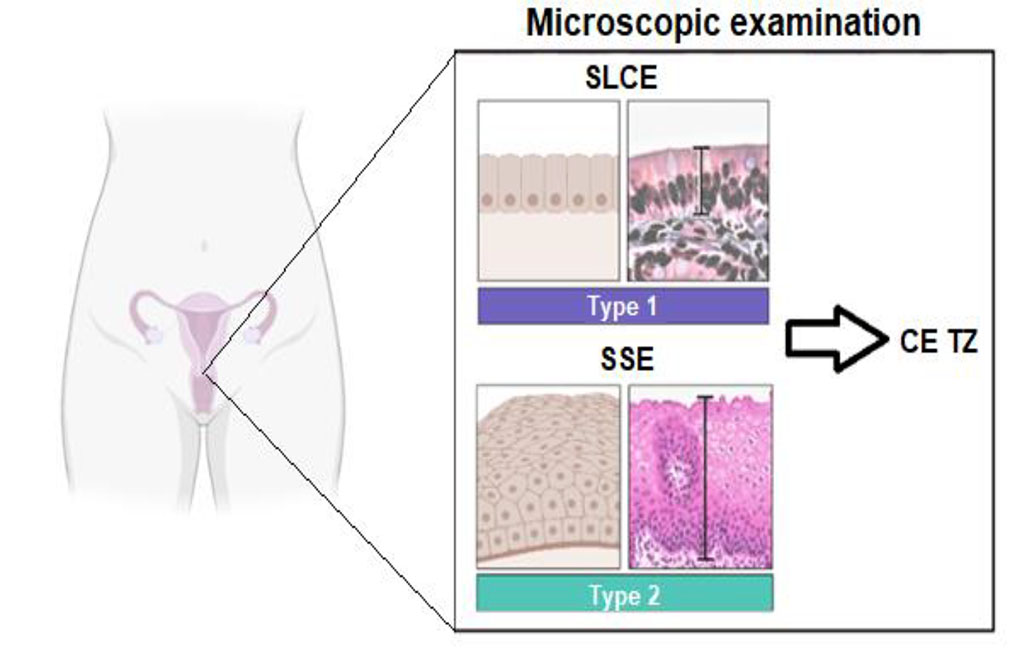
Figure 1. Scheme of the cervical epithelium. Note: SSE - stratified squamous epithelium of the exocervix; CE TZ - columnar epithelium of the transformation zone; SLCE - single-layered cylindrical epithelium of the endocervix
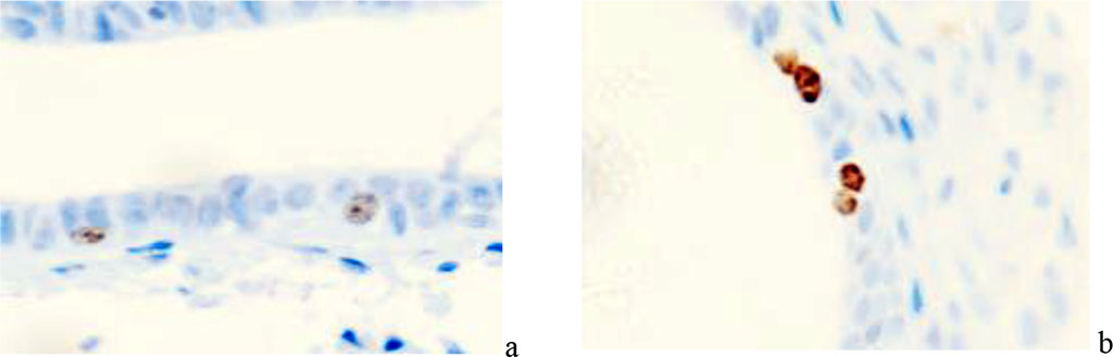
Figure 2. A 63-year-old woman. Localization of Ki67 gene protein in the structures of the cervical mucosa in normal postmenopause. Immunohistochemistry with hematoxylin dyeing. Microphoto. Eq. x400
Thus, we observed that in postmenopausal women there is an increase in receptors for progesterone, a decrease in estrogen levels is associated with a decrease in receptors for estrogens and a sharp decrease in proliferative activity in the epithelial lamina of the CM (Fig. 3).
The number of Ki67-positive cells in the pathologic course during postmenopause increases to a greater extent in the exocervical SMA compared to the norm, which can be attributed to pathogen contamination and adaptive sloughing of the epithelium on the epithelial surface, these data for norm and pathology are presented in Table 1, 2.
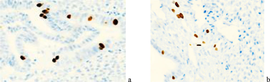
Figure 3. Localization of Ki67 gene protein in the structures of the cervical mucosa in the presence of polyps in the cervical mucosa. Microphoto. Immunohistochemistry. Eq. x200
The number of cells positive for Ki-67 during the pathologic process in postmenopause increases, especially in the epithelium of the exocervical epithelium of the SMA compared to the norm. This phenomenon can be attributed to colonization of this area by pathogens and adaptive desquamation of the epithelium on the tissue surface. The corresponding data on norm and pathology are presented in Tables 1 and 2.
Table 1. Evaluation of the location and quantification of Ki-67-positive cell activity in the CM of patients with normal course of postmenopausal period
| CM structures control | Average Ki-67 gene activity (number of mitoses per 100 cells in %) | ||||
| 1 year PMP | 2 year PMP | 3 year PMP | 4 year PMP | 5 year PMP | |
| EExocervix | 3,2 | 2,4 | 2 | 2,9 | 1,2 |
| Ttransformation zone | 2,4 | 2,4 | 2,1 | 2,6 | 1,1 |
| EEndocervix | 1,8 | 2,4 | 1,9 | 1,5 | 1,1 |
| TTotal | 7,4 | 7,2 | 6 | 7 | 3,4 |
Table 2. Evaluation of location and quantitative characterization of Ki-67 - positive cells activity in CM of patients with pathological course of postmenopausal period
| CM structures in the observation group | Average activity of Ki-67 gene (number of mitoses per 100 cells in %) | ||||
| 1 year PMP | 2 year PMP | 3 year PMP | 4 year PMP | 5 year PMP | |
| EExocervix | 4,2 | 2,9 | 2,5 | 2,6 | 1,7 |
| TTransformation zone | 3.2 | 2,5 | 2,2 | 1,8 | 1,3 |
| EEndocervix | 2,5 | 2,4 | 1,9 | 1,6 | 0,8 |
| TTotal | 9,9 | 7,8 | 6,6 | 6 | 3,8 |
Based on the data, it was determined that there were differences in the exocervix at any period (average over the next 5 years after the onset of expression at 2.34% for normal vs. 2.78% for pathology), and in the transformation zone (2.4% for normal vs. 3.2% for pathology) and endocervix (1. 8% at normal vs. 2.5% at pathology) in the first year of postmenopause as a function of PMP normality and increased mean Ki-67 expression level at pathology; no differences were observed in the transformation zone and endocervix in subsequent years after the onset of postmenopause with respect to PMP progression.
Conclusion. In the course of the study, it was found that different sections of the BMS have peculiarities of regenerative potential, characterized by the activity of the Ki-67 gene, which varies in the age dynamics of the examined groups. The highest activity is observed in the exocervix area, the lowest values in the transformation zone and the lowest values are observed in the endocervix area.
The concept and design of the study - D.P. Puga, G.V. Reva, I.V. Reva
Collection and processing of material - D.P. Puga, G.V. Reva, I.V. Reva
Text writing - D.P. Puga, G.V. Reva, B.O. Shcheglov
Editing - I.P. Koval, M.B. Khamoshina, V.V. Usov, K.V. Stegniy, S.N. Shcheglova

|
||