- Home
- About the Journal
- Peer Review
- Editorial Board
- For Authors
- Reviewer Recognition
- Archive
- Contact
- Impressum
- EWG e.V.
The aim of the work was to evaluate the dielectric properties of human tissues in ex vivo conditions. The study was performed on 68 tissue samples, 20 of which were fragments of muscle tissue, 17 – fatty, 11 – fibrous and 20 – a full-layer skin flap. The analysis of the dielectric properties of tissue fragments was carried out by the method of near-field resonant microwave sounding using a special software and hardware complex. The study made it possible to verify the presence of pronounced differences in the dielectric properties of tissues depending on their histotype. It was revealed that adipose tissue has the highest level of dielectric permittivity, and fibrous tissue has the lowest one. Muscle tissue and a full-layered skin flap occupy an intermediate position between them. These data are fundamentally important for the creation of standards for the dielectric characteristics of biological objects and the further development of near-field resonant microwave sensing technology as an innovative medical imaging technology.
Keywords: near-field microwave sensing, biological tissue, dielectric properties
For a long time, various research groups have been trying to study the state and characteristics of body tissues by their dielectric properties [1-4, 7, 8]. At the same time, there is still no unified methodological base and methodological apparatus for assessing the dielectric parameters of biological objects [6-8]. In the literature, there are different opinions about the optimal probing frequencies (from tens of MHz to 100 GHz), as well as the most informative method of probing (active or passive) [3, 6, 7]. As a result, integrative reference physiological intervals for tissues of various histotypes have not been formed. In our opinion, the use of remote intraoperative biological material for this purpose is an assumed option for the analysis of individual tissue types [6].
Taking into account the above, the aim of the work was to evaluate the dielectric properties of human tissues in ex vivo conditions.
The study was performed on 68 tissue samples, 20 of which were fragments of muscle tissue, 17 – fatty, 11 – fibrous and 20 – a full-layer skin flap. The thickness of each fragment in one of the measurements was at least 5 mm (depth of sounding). All samples were analyzed within no more than 1 hour after receipt. All patients whose biomaterial was analyzed gave informed consent to participate in the study.
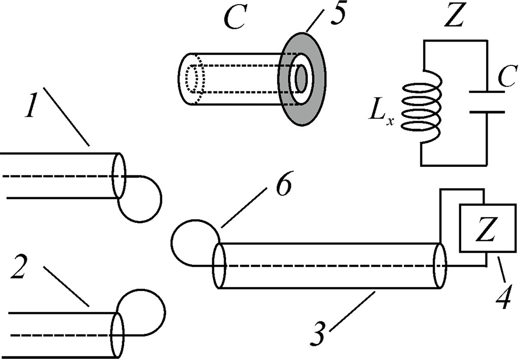
Figure 1. Electrodynamic model of resonance near-field measuring system: 1 – exiting line, 2 – receiving line, 3 – resonator, 4 – load of resonator, 5 – near-field antenna as cylindrical capacitor, 6 – magnetic loop of resonator
The analysis of the dielectric properties of tissue fragments was carried out by the method of near-field resonant microwave sounding using a special software and hardware complex of our own design (Fig. 1) [5, 6]. For this experiment, a sensor with a sounding depth of 5 mm was used to evaluate the dielectric permittivity of fragments. The study of each sample was performed at 3 different points, then the average value for them was calculated. The probe frequency of the sensor was 789 MHz.
All biological tissue samples were obtained after the patients signed informed consent. The study was approved by Local Ethic Committee of Privolzhsky Research Medical University.
Statistical processing of the obtained results was performed using the program Statistica 6.1 for Windows.
It was found that there are significant variations in their dielectric parameters between fragments of different tissues (Fig. 2).
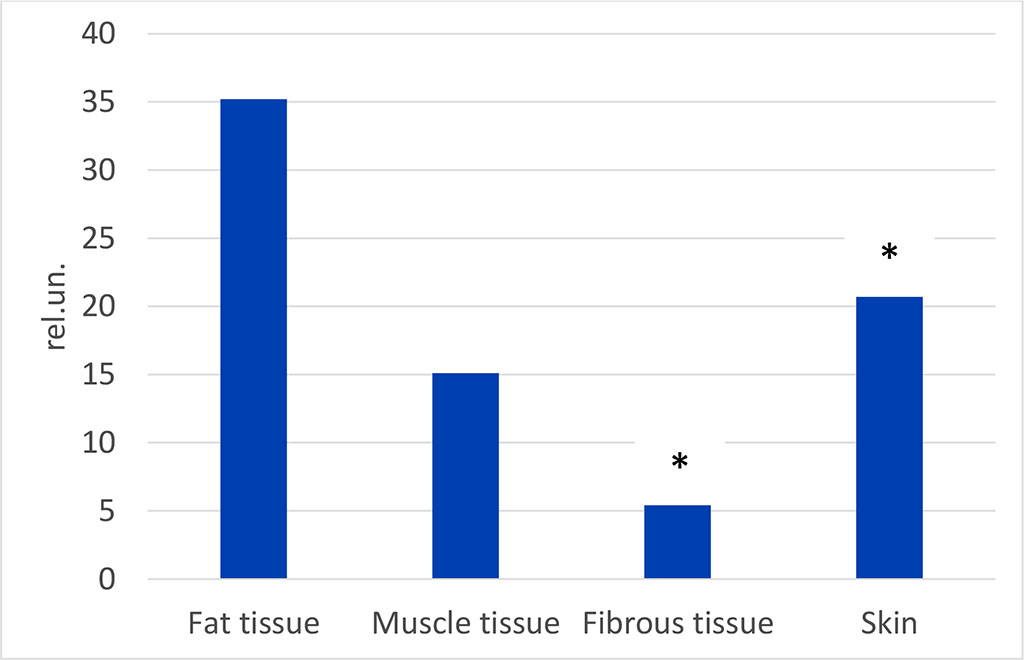
Figure 2. The level of dielectric permittivity of biological samples of different tissues («*» - difference to fat tissue is statisitcally valied, p<0.05)
Thus, adipose tissue has the greatest dielectric permeability, and the remaining histotypes demonstrate a significantly lower level of the parameter. In particular, the permeability of muscle tissue is 2.33 times characteristic of fatty tissue (p<0.01), and fibrous tissue is 6.52 times lower (p<0.001). At the same time, a full-layered skin flap occupies an intermediate position (between these types of tissues, being 1.70 times smaller than that identified for adipose tissue (p<0.01).
According to classical concepts, the level of dielectric permittivity directly depends on the water content in the analyzed object [3, 4, 6]. This explains the discovered features of the dielectric properties of tissues of various histotypes.
The study made it possible to verify the presence of pronounced differences in the dielectric properties of tissues depending on their histotype. It was revealed that adipose tissue has the highest level of dielectric permittivity, and fibrous tissue has the lowest. Muscle tissue and a full-layered skin flap occupy an intermediate position between them. These data are fundamentally important for the creation of standards for the dielectric characteristics of biological objects and the further development of near-field resonant microwave sensing technology as an innovative medical imaging technology.
This study was particularly supported by Russian Science Foundation (project No 22-25-00652).

|
||