- Home
- About the Journal
- Peer Review
- Editorial Board
- For Authors
- Reviewer Recognition
- Archive
- Contact
- Impressum
- EWG e.V.
Aim: to study the morphometric characteristics of the mandibular condylar processes to identify patterns and their classification.
Materials and Methods: a study of 49 skeletonized and certified mandibular preparations was performed using a caliper and a depth gauge. During the study of the material, the forms and the main morphometric parameters of the condylar processes were determined and analyzed. Statistical data processing was carried out using the Microsoft Excel program.
Results: The study of certified biological material yielded the main morphometric and morphofunctional parameters of the mandibular condylar processes. We revealed characteristic differences in the anatomical structures of the studied preparations. The main variants and types of combinations of forms of condylar processes were singled out: oval, hook-shaped, diamond-shaped, L-shaped. The lowest asymmetry index is characteristic of a combination of oval and diamond–shaped forms - 2.44%., and the highest asymmetry index is characteristic of diamond–shaped and L-shaped forms of condylar processes - 7.13%.
Conclusion: The presented data can help practitioners to more accurately determine the target point for Gow-Gates anesthesia; to take into account the nature of the influence of structural changes in the joint on other elements of the dental apparatus, thus preventing functional diseases of the temporomandibular joint. The methodology of studying the main morphometric parameters on skeletonized preparations of the lower jaws can be adapted for the examination of living persons using diagnostic radiology.
Keywords: morphometric examination, condylar process, Gow-Gates anesthesia, mandible
The condylar processes of the lower jaw are important links of the dental apparatus. Pathological processes that occur directly in the temporomandibular joint, the surrounding bone and soft tissue structures lead to disruption in the myodynamic balance and serious dysfunction in controlling jaw movements, such as opening the mouth, chewing food and external respiration. The nature of the influence of structural disorders of the condylar processes on other areas of the dental apparatus and its consequences on the features and effectiveness of therapeutic manipulations is not fully understood.
Taking into account the clinical significance and relevance of this issue, morphometric characteristics of the condylar processes of the mandible were studied in order to identify patterns and their classification.
Morphological and morphometric features of the temporomandibular joint are formed under the influence of pathological processes in the prenatal and postnatal periods of human development. [12]
In the postnatal period, during its development the lower jaw is exposed to various factors: age-related changes, traumatic injuries, the development of inflammatory processes, changes in the adequate distribution of masticatory pressure, changes in occlusal relationships, disorganization of masticatory muscle groups, etc. These factors cause structural changes in the lower jaw, which affects the nature of muscle attachment.[22]
Individual variability of anatomical and topographic features of the condylar processes of the mandible, arising under the influence of both congenital and acquired factors, is one of the causes of complications after local anesthesia, as well as the cause of pain dysfunction of the TMJ, disorders of myodynamic balance and functions of chewing, breathing and external respiration.
Previously, various authors have tried to classify the shape of the condylar processes of the mandible [5, 7, 13, 15, 21, 25]. Orthopantomographic systematization, which was used by Anisuzzaman M. M. et al., can be considered the most successful. (2019) in his work on the study of the morphology of the condylar processes of the mandible, which, according to this classification, can have the shape of an oval, a bird's beak, a rhombus and a curved finger. [2]
The morphology of the condylar process is of great importance for applied medicine and diagnostic radiology as well as for planning surgical interventions, blockades, and providing emergency care in maxillofacial surgery [1, 3, 6, 8, 16, 19, 20].
Awareness of TMJ pathologies of various etiologies (ankylosis, osteoarthritis, osteoarthritis, malignant neoplasms, callus, etc.) that reduce the quality of intraoral dental manipulations (for example, IANB) due to the restriction of mouth opening is also of great practical importance in the clinical activity of a dentist. [9, 13, 24]
Consequently, knowledge of the morphometric characteristics of the condylar process helps avoiding the development of dental complications, such as: mechanical damage to the pterygoid muscle and contracture of the mandible, injury to the inferior alveolar nerve and vessels with the formation of a hematoma, the ingress of anesthetic into the bloodstream, etc. [10, 11, 17, 23].
A study of the lower jaws from 49 corpses (male – 32, female – 17) was conducted. The age of the species was in the range of 72.67±11.47 years. The study was conducted at the Department of Operative Surgery and Topographic Anatomy, Sklifosovsky Institute of Clinical Medicine and I.M. Sechenov First Moscow State Medical University (The Sechenov University). This study was approved by the Ethics Committee of The Sechenov University (Protocol no.: 02-23 of 01/26/2023). Preparations of the mandible with the presence of congenital defects and deformities, as well as with a violation of the safety of the preparations, i.e. the anatomical integrity of the condylar processes, branches and the body of the mandible, were excluded from the study.
During the study of the material, the forms and the main morphometric parameters of the condylar processes were determined and analyzed.
Statistical data processing was carried out using the Microsoft Excel program. The sample size is determined by calculating the formula:
Where n is the volume of the sample population; N is the size of the general population; Z is the normalized deviation determined based on the selected confidence level; p is the variation found for the sample; q=1–p; e is the marginal sampling error (for the proportion of the trait, the confidence interval ("error" ± %).
For the general population of 49 skeletonized and certified lower jaw preparations with a confidence level of 95% and a maximum permissible error of 5%, the volume of the sample population was 44.
The arithmetic mean, standard deviation, arithmetic mean error, Student's t-test and asymmetry index are determined. The differences were considered statistically significant at p<0.05.
The following parameters were analyzed: the thickness of the base of the condylar process, the height of the condylar process, the length of the base of the condylar process, the width of the condylar process in cross-section, the width of the condylar process in sagittal section, the shape of the condylar process. (Fig. 1, 2), the distance from the head of the lower jaw to the mandibular opening. Two main samples were formed: depending on the gender and the shape of the head of the lower jaw. The distribution of the mandibles according to the shape of the condylar processes was carried out taking into account the gender in accordance with the classification proposed by us.
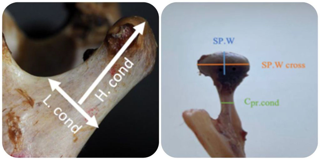
Figure 1. Measurement methodology: H.cond - height of the condylar process; L.cond – length of the base of the condylar process; Cpr.cond – thickness of the base of the condylar process; SP.W is the width of the condylar process in the sagittal section; SP.Wcross transversely is the width of the condylar process in cross section.
Statistical data processing was carried out on the basis of the Microsoft Excel program using descriptive statistics methods (determination of the arithmetic mean, standard deviation, average error of the arithmetic mean, Student's t-test, asymmetry index). The differences were considered statistically significant at p <0.05.
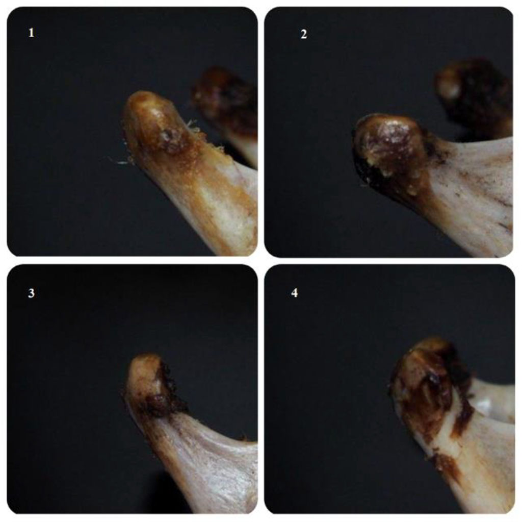
Figure 2. The shapes of the condylar processes of the lower jaw. 1. The condylar process has an oval shape; 2. The condylar process has a diamond–shaped shape; 3. The condylar process has the shape of a hook; 4. The condylar process has an L-shaped shape.
In the course of the study, two main theses were formulated:
According to the results of morphometric measurements of the condyle processes of the lower jaw, the results were statistically processed. These results of the study are presented in the tables 1, 2, 3, 4, 5.
Table 1. Statistical analysis of morphometric measurements of the condylar processes in men
| thickness of the base of the condylar process | height of the condylar process | length of the base of the condyle process | width of the condylar process in cross section | width of the condylar process in sagittal section | ||||||
| Side | R | L | R | L | R | L | R | L | R | L |
| arithmetic mean | 0,69 | 0,72 | 2,46 | 1,88 | 1,54 | 1,51 | 2,03 | 2,02 | 0,86 | 0,90 |
| standard deviation | 0,14 | 0,14 | 2,47 | 0,35 | 0,22 | 0,23 | 0,20 | 0,26 | 0,18 | 0,19 |
| the average error of the arithmetic mean | 0,03 | 0,03 | 0,47 | 0,07 | 0,04 | 0,04 | 0,04 | 0,05 | 0,03 | 0,04 |
| М1 | 0,74 | 0,77 | 3,41 | 2,02 | 1,63 | 1,60 | 2,11 | 2,12 | 0,93 | 0,98 |
| М2 | 0,64 | 0,66 | 1,50 | 1,75 | 1,46 | 1,42 | 1,96 | 1,92 | 0,79 | 0,83 |
Notes to Table 1: R is the right condylar process, L is the left condylar process, M1 and M2 are the confidence limits of the arithmetic mean.
Table 2. Statistical analysis of morphometric measurements of the condylar processes in women
| thickness of the base of the condylar process | height of the condylar process | length of the base of the condylar process | width of the condylar process in cross section | width of the condylar process in sagittal section | ||||||
| Side | R | L | R | L | R | L | R | L | R | L |
| arithmetic mean | 0,67 | 0,68 | 1,81 | 1,73 | 1,45 | 1,39 | 1,89 | 1,93 | 0,81 | 0,84 |
| standard deviation | 0,13 | 0,13 | 0,34 | 0,33 | 0,13 | 0,16 | 0,21 | 0,23 | 0,12 | 0,15 |
| the average error of the arithmetic mean | 0,03 | 0,03 | 0,08 | 0,08 | 0,03 | 0,04 | 0,05 | 0,05 | 0,03 | 0,03 |
| М1 | 0,73 | 0,74 | 1,97 | 1,88 | 1,51 | 1,47 | 1,99 | 2,04 | 0,86 | 0,91 |
| М2 | 0,60 | 0,62 | 1,64 | 1,57 | 1,39 | 1,31 | 1,79 | 1,82 | 0,75 | 0,76 |
Notes to Table 2: R is the right condylar process, L is the left condylar process, M1 and M2 are the confidence limits of the arithmetic mean.
Table 3. Definition of the Student's t-test.
| Student's t-criterion | thickness of the base of the condylar process | height of the condylar process | length of the base of the condylar process | width of the condylar process in cross section | width of the condylar process in sagittal section | |||||
| R | L | R | L | R | L | R | L | R | L | |
| 0,58 | 0,97 | 1,38 | 1,58 | 1,79 | 2,04 | 2,39 | 1,22 | 1,31 | 1,34 | |
Notes to Table 3: R – right condylar process, L – left condylar process
According to the calculation of the Student's t-test presented in Table 3, it follows that the statement about the presence of larger morphometric indicators of condylar processes in men is reliable. The results obtained are consistent with the studies of other researchers [4, 14].
In 49 skeletonized preparations of the mandible, no statistically significant differences in the shape of the condylar process, depending on whether the drug belongs to one of the sexes, were found. The most common forms of the mandibular head among the studied preparations were the forms: L-shaped and in the form of a diamond. At the same time, on the same preparation of the lower jaw, the shapes of the condyle processes on the right and left could differ. We have identified the following combinations of forms of condylar processes on the right and left on the lower jaw preparations:
After determining the combination of the shapes of the condylar processes, measurements were made of the distance from the heads of the condylar processes to the mandibular foramen on the right and left. According to the measurement, the following results were obtained:
Table 4. Average values of the distance from the condylar processes to the mandibular foramen on preparations of the mandible in men
| preparations of the mandible with an oval shape condylar process | preparations of the mandible with a hook-shaped condylar process | preparations of the mandible with a diamond-shaped condylar process | preparations of the mandible with an L-shaped condylar process | |||||
| Side | R | L | R | L | R | L | R | L |
| M gen. | 3,77±0,56 | 3,98±0,35 | 4,02±0,42 | 3,68±0,45 | 3,98±0,33 | 4,04±0,39 | 4,00±0,20 | 4,05±0,14 |
Note to Table 4: R is the right condylar process, L is the left condylar process, M gen is the confidence limits of the arithmetic mean distance from the head of the mandible to the mandibular foramen.
Table 5: Average values of the distance from the condylar processes to the mandibular foramen on preparations of the lower jaw in women
| preparations of the mandible with an oval shape condylar process | preparations of the mandible with a hook-shaped condylar process | preparations of the mandible with a diamond-shaped condylar process | preparations of the mandible with an L-shaped condylar process | ||||
| Side | R | L | R | R | L | R | L |
| M gen. | 3,73±0,41 | 3,76±0,73 | 3,92±6,60 | 3,73±0,22 | 3,56±0,51 | 3,48±0,39 | 3,61±0,70 |
Note to the table: R – right condylar process, L – left condylar process, M gen. – confidence limits of the arithmetic mean distance from the head of the mandible to the mandibular opening.
In the group of mandibular preparations, there are no mandibular preparations with a hook-shaped condylar process on the left, due to the presence of only one mandible with such a condylar process shape, which did not allow statistical calculations to determine the arithmetic mean.
Analyzing the data presented in Tables 4 and 5, it follows that the distance from the mandibular heads to the mandibular opening in men is slightly greater than in women. Clinically, this distance on the inner surface of the lower jaw determines the level of needle insertion with various methods of local anesthesia. For example, for local anesthesia of the mandible, the target point of which is the inner surface of the lower jaw branch in the area slightly receding posteriorly from the entrance to the mandibular foramen.
To clarify the validity of the thesis about the influence of the shapes of the condylar process on the development of structural and topographic changes in the anatomical formations of the mandible, we calculated the asymmetry index for each combination of the shapes of the condylar processes on the right and left. In the course of the calculations, the following results were obtained: the asymmetry index for the combination of 2 oval forms of condylar processes on the right and left was 3.04%. The index of asymmetry in the combination of an oval shape with a hook-shaped condyle was - 4.05%. For a combination of oval and diamond-shaped shapes - 2.44%. For a combination of an oval shape with an L-shaped condyle – 4.64%. When combining two hook-shaped forms of the condylar process, the asymmetry index was 3.77%. When the diamond-shaped condyle was combined with a hook-shaped shape, the asymmetry index was 5.42%. For the hook–shaped and L-shaped forms of the condyle process, the asymmetry index is -6.62%. For a combination of two diamond-shaped forms of the condylar process, the asymmetry index was 3.29%. For the diamond-shaped condyle and the L–shaped condyle, the index was 7.13%. Two L-shaped forms of the condyle are characterized by an asymmetry index equal to 4.34%.
Based on the data obtained, it follows that the lowest indices of the asymmetry index are mainly characteristic for preparations of the lower jaws with two identical forms of condyle processes on the right and left. However, the lowest indicator was found in lower jaw preparations with a combination of oval and diamond-shaped condylar process. Higher indices of the asymmetry index are characteristic of lower jaw preparations with various forms of condylar processes. This fact confirms the existence of a relationship between the change in the shape of the mandibular head and the different topographic location of the mandibular opening, which is important for choosing an effective anesthetic technique of the mandible.
Our data shows that the lowest indices of the asymmetry index are mainly characteristic of preparations of the lower jaws with two identical forms of condylar processes on the right and left. However, the lowest indicator was found in preparations of the lower jaw with a combination of oval and diamond-shaped condyle. Higher indices of the asymmetry index are characteristic of mandibular preparations with various forms of condylar processes. This fact confirms the existence of a relationship between the change in the shape of the condylar processes and the different topographic location of the mandibular opening, which is important for the effective anesthetic method of the mandible.
In the course of the study, we established differences in the shapes of the condylar processes and the different topographic location of the mandibular foramen with the revealed combinations of the shapes of the condyle processes. Modern classifications of the forms of condylar processes were studied. [2] The disadvantage of the classification by Anisuzzaman M. M. et al. (2019) is dealt with the subjectivity of the vision of the form, which is based on a figurative interpretation of the X-ray image in 2D format. The lifetime mechanisms of changes in the shapes of the condylar processes in individuals on each side and their effect on the change in the relative position of the mandibular openings on the lower jaw require further study. Assessing possible causes of morphometric and morphological changes in the condylar processes of the mandible, we assume the presence of occlusive disorders, unilateral overload of the condylar processes of the mandible, functional disorders of the masticatory muscles, etc. The average age of patients 72.67 ±11.47 years, physiological bone resorption, the formation of senile bite – these factors influence the changes in the topographic location of the mandibular foramen. The picture will be clearer when studying the lower jaws in young and middle-aged people using radiological examinations and morphometric measurements on lower jaw preparations.
In this study, we found a pattern of changes in the topographic location of the mandibular opening with different forms of condylar processes, both symmetrical on both sides of the mandible, and differing in shape on one side and on the other. In connection with the discovered regularity, we believe that in order to ensure the effectiveness and safety of the technique for mandibular conduction anesthesia, morphometric features of the elements of the condyle of the mandible should be taken into account.

|
||