- Home
- About the Journal
- Peer Review
- Editorial Board
- For Authors
- Reviewer Recognition
- Archive
- Contact
- Impressum
- EWG e.V.
Cite as: Archiv EuroMedica. 2022. 12; 3: e1. DOI 10.35630/2199-885X/2022/12/3.4
The aim of this study was to estimate changes of tissue dielectric parameters under the action of high temperature. We fixed the shifts of dielectric properties of biological tissue samples, associated with short-time influence of high temperature (600C, 5 min.) in vitro. The dielectric properties of tissues (permittivity - ε and conductivity - σ) were determined using original software and hardware complex for near-field resonant microwave sensing developed at the Institute of Applied Physics of the RAS. The study of these parameters was performed at a depth of 5 mm. It was found that short processing of a biological sample in moderate hyperthermia (600C) leads to significant change of dielectric characteristics of the tissue. This is manifested in a significant decrease in the dielectric permittivity (in 2.48 times for an intact specimen) and conductivity (on 25.3%) of the studied biological sample, due to its dehydration. It was shown that used regimen of heating decreases the permittivity and conductivity of tissue specimen that is associated with changes of its hydratation.
Keywords: near-field microwave sensing, biological tissue, dielectric properties
Currently, there are still a significant number of diagnostic difficulties associated with clarifying the boundaries of the burn lesion, the viability of tissues in the parawound zone, the uniformity of the wound etc. [1, 3, 4, 6, 7]. A separate question is the verification of the depth of skin and subcutaneous structures [2, 3, 4, 6, 7]. To solve this set of problems, in addition to the empirical approach prevailing in real clinical practice, the possibilities of thermal IR-imaging were studied [2, 9]. It is shown that in a number of situations this technology is informative, but it allows us to judge only the state of the skin surface and the nearest underlying structures. Modern versions of ultrasound examination, which have high information content and resolution in other pathologies, do not allow achieving the desired contrast in relation to thermal trauma [1, 2, 7]. An additional complicating circumstance in tissue visualization in combustiology is the presence of a physical barrier between the sensor and the skin surface (temporary and permanent wound coverings), which is not always possible to eliminate for diagnostic manipulations (for example, when using biosurfaces containing a matrix with stem cells) [1, 7]. This is an obstacle for most of the methods of studying the subsurface structure, integumentary tissues, in particular, for ultrasound research. Therefore, it is necessary to search an alternate method for andor to test fundamentally different technologies for assessing the deep characteristics of the burn wound and the parawound zone [4, 6, 7].
In this aspect, attention is drawn to the relatively recent method of near-field resonant microwave profiling in biomedicine, based on the study of the dielectric properties of tissues (permittivity and conductivity) [2, 5, 8]. On the other hand, its possibilities for combustiology are only beginning to open [2].
In this regard, the aim of the study was to assess the nature of shifts in the dielectric parameters of the tissue under the influence of high temperature.
The current experiment was performed on equal samples of palmar aponeurosis tissue, which was removed intraoperatively (n=8). The thermal exposure was modeled by placing of tissue fragments in a thermostat (processing time - 5 min., temperature - 600C). The duration of exposure thermal modification was 5 minutes.
The dielectric properties of tissues (permittivity - ε and conductivity - σ) were determined using original software and hardware complex for near-field resonant microwave sensing [2, 5]. The study of these parameters was performed at a depth of 5 mm, approximately comparable to the thickness of the analyzed tissue samples. Study of each piece of tissue was carried out twice – before and after thermal modification (at the conclusion of the exhibition period).
All biological tissue samples were obtained after the patients signed informed consent. The study was approved by Local Ethic Committee of Privolzhsky Research Medical University.
Statistical processing of the obtained results was performed using the program Statistica 6.1 for Windows.
Results of the study revealed that as a result of thermal modification, the dielectric characteristics of biological tissue are significantly transformed (fig. 1-2). This might be due to the fact that these are significantly depend on the content of water in the studied object [3, 5, 6, 8], and when heated, there is a significant decrease in tissue hydration. Thus, after drying, a decrease in the dielectric permittivity of the samples was observed by 2.48 times compared to the initial level of the indicator (fig. 1; p<0.05).
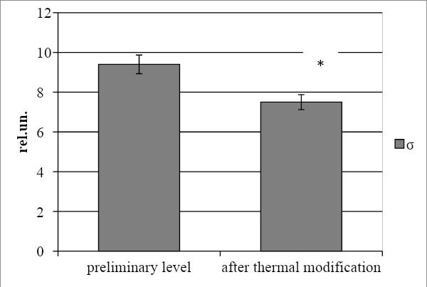
Figure 1. The dynamics of dielectric permittivity of biological samples under thermal modification; («*» - level of statistical value (p<0.05)
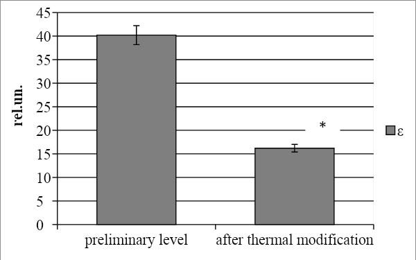
Figure 2. The dynamics of dielectric conductivity of biological samples under thermal modification; («*» - level of statistical value (p<0.05)
Similar dynamics of the parameter were reported in previous studies, which included a comparative assessment of the dielectric properties of the skin and subcutaneous structures in healthy Wistar rats with model thermal trauma [2, 6]. This confirms the diagnostic informativeness of the analysis of the dielectric permittivity of tissues under the influence of high temperatures, including thermal burns.
A similar but less pronounced trend was found for the conductivity of the tissue sample (fig. 2). It was found that after thermal modification, the value of this indicator decreases by 25.3% relative to the intact state (p<0.05). This indicates a violation of the physiological structure of the tissue and indirectly indicates metabolic shifts in it, which cause changes in the ionic composition of the intercellular substance, its osmolarity and other physical and chemical characteristics [3, 4, 9]. This fact can be used as an experimental justification for the possibility of detecting the boundaries of thermal tissue damage that occurs in real burns.
It was found that short processing of a biological sample in moderate hyperthermia (60℃) leads to significant change of dielectric characteristics of the tissue. This is manifested in a significant decrease in the dielectric permittivity and conductivity of the studied biological sample, due to its dehydration, which occurs under the selected mode of thermal modification. It should be emphasized that these data obtained in the ex vivo experiment are fully consistent with the results of microwave research of experimental burn wound tissues [2, 6]. This confirms the diagnostic informativity of the technology.

|
||