- Home
- About the Journal
- Peer Review
- Editorial Board
- For Authors
- Reviewer Recognition
- Archive
- Contact
- Impressum
- EWG e.V.
Cite as: Archiv EuroMedica. 2022. 12; 3: e1. DOI 10.35630/2199-885X/2022/12/3.14
The authors set a goal to improve the results of treatment in patients with staple line failure after bariatric surgery using various endoscopic techniques. Based on four clinical cases an algorithm for the sequence of actions was created. The authors used vacuum aspiration, stent placement, as well as sealing the organ defect with bioglue. In all four cases, it was possible to successfully cope with the staple line failure and prevent the progression of peritonitis. As a result of the study, the authors conclude that several endoscopic treatments can be considered in tandem or in combination. The treatment of this category of patients should be interdisciplinary and include an adequate timing.
Keywords: staple line failure, bariatric surgery, mediastinal fistula, surgical complications, endoscopic treatment
Suture failure is a serious complication during operations on the hollow organs of the digestive tract, associated with a high duration of inpatient treatment and mortality. In a series of early reports dating back to the 1990s, the incidence of esophageal anastomosis leakage after gastrectomy and proximal gastric resection was as high as 30%. Publications of recent years show a clear positive trend in the decline in the incidence of failure, which is a reflection of the unconditional progress in surgical technique and instrumental improvements [1,2].
The choice of management tactics for each patient is individual. Endoscopic techniques have played a significant role in the diagnosis and treatment of this complication. Endoscopy allows diagnosing the presence of a defect, as well as assessing its location and size. In recent decades, a number of methods have been developed for endoscopic treatment of anastomosis and suture failure in the digestive tract, which have a number of advantages over surgical and conservative management of this group of patients. Despite the accumulated experience, there is currently no unified systematic approach to the use of endoscopic methods for closing anastomotic defects [3,4,5].
The treatment strategy for a patient with failed sutures of the upper digestive tract is aimed at the simultaneous solution of several problems: sanitation of the cavity and drainage of the mediastinum, providing nutritional support, suture plication, prevention and treatment of purulent complications [6,7,8].
Purpose of the study: To optimize the results of treatment in patients with staple line failure after bariatric surgery using various endoscopic techniques.
Initially, it is necessary to ensure adequate sanitation and drainage of the abscess, with the introduction of antiseptic solutions and agents. Usually this problem is solved surgically, depending on the nature of the previously performed surgical intervention and the type of surgical access. In a number of cases, adequate sanitation can be achieved by installing drains under ultrasound or computed tomography control, and even using flexible endoscopes through the formed hole in the anastomosis.
Recently, endoscopic vacuum therapy has been used to ensure adequate sanitation and drainage of the purulent cavity. The operation is performed with the patient lying on his back. To ensure the stability of the respiratory function at all stages of the intervention, orotracheal intubation was performed, followed by artificial lung ventilation. To reduce the risk of possible complications, an intervention was performed using carbon dioxide. During the initial study, the level of location of the anastomotic leak was noted, which was measured in centimeters from the incisors. The size of the leak and the local state of the tissues, the degree and severity of inflammatory and regenerative changes were determined. When technically possible and the patient's condition was stable, the leakage cavity in the mediastinum or abdominal cavity, communicating with the failure site, and the fistulous tract were inspected. Their sizes, the state of the surrounding tissues were assessed with the determination of the severity of inflammatory changes, as well as the presence of sequesters. If necessary, to specify the identified changes, assess the configuration of the cavity, diagnose latent streaks, and determine the adequacy of the drainage, the cavity was contrasted using a water-soluble contrast. Primary sanitation of the cavity with the removal of fibrin, saliva and intestinal contents and necrotic tissues was carried out by abundant washing with sterile solutions with delicate aspiration of the contents, excluding damage to adjacent anatomical structures and, above all, blood vessels. Also, during the initial study, the technical feasibility of inserting a porous polyurethane system into the cavity lumen without the risk of additional anastomotic injury was determined. Subsequently, the endoscope was passed below the level of the anastomosis at a distance of at least 50 cm with the installation of a nasoenteric nutritional tube.
In accordance with the information received about the size of the leak and the lumen of the esophagus, the final decision was made on the nature of the placement of the porous system - inside the esophagus or inside the cavity. Guided by the decision made, the primary sponge system was cut out from the polyurethane sponge plate using Cooper's scissors and its shape was modeled to a cylindrical one. When the sponge was located intraesophageally, its average diameter corresponded to the diameter of the esophagus at the level of the anastomosis or was slightly smaller than the latter. When located inside the cavity, the diameter of the spongy cylinder corresponded to the size of the defect, but not more than the diameter of the esophagus. The length of the simulated spongy implant with intraesophageal location was determined by the length of the defect with overlapping of the wound edges by 3 cm on both sides and most often was 6-8 cm or more. Fig.
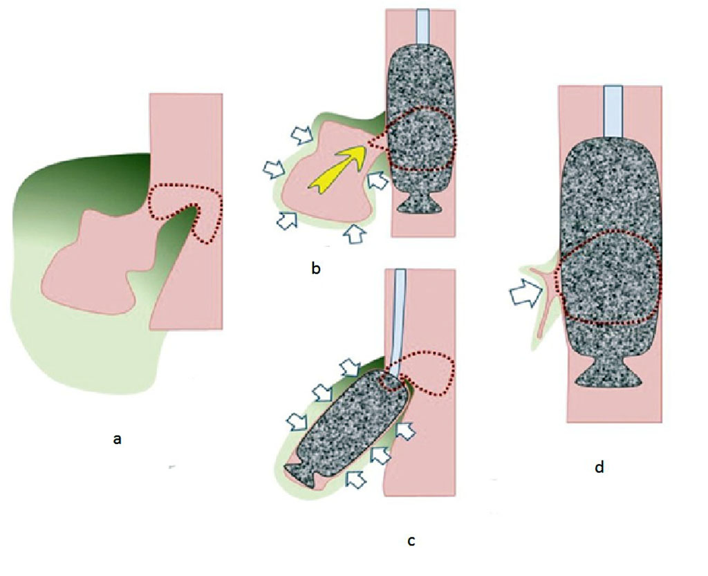
Fig. Schematic arrangement of the vacuum aspiration system. a - anastomotic failure (marked by a dotted line) with the presence of a cavity (red color) and inflammatory changes in the surrounding tissues (green color); b - installation of a vacuum aspiration system in the lumen of the esophagus. The yellow arrow indicates the direction of evacuation of the contents from the cavity. White arrows indicate the direction of retraction of adjacent tissues under the influence of negative pressure created in the porous system. The dotted line marks the configuration of the anastomotic line with the vacuum aspiration system installed; c - installation of a vacuum aspiration system in the cavity near the site of the leak. The designations are the same; d - changes in the anastomosis and surrounding tissues at the stages of treatment. The designations are the same.
A porous sponge mounted on a gastric tube was placed parallel to the endoscope and immediately before insertion was grasped by the right hand together with the inserted part of the apparatus as a single system. The subsequent establishment of the system was carried out under visual control through the endoscope. To reduce the effort during the system and overcome the natural resistance in the area of anatomical narrowing, it is advisable to use an endoscope of increased rigidity, moisten a porous sponge with water or a water-soluble gel, slightly unbend the head in the cervical region in order to straighten the bend in the pharyngeal-esophageal region.
The level of location of the porous system was determined under direct visual control through the endoscope. Taking into account that the sponge is located outside the field of view of the apparatus, the latter was passed distally to the level of the defect into the anastomosed intestine or stomach to a depth of 4–5 cm. Subsequently, the towel clip inserted through the endoscope channel and fixing the porous system at the distal end by the loop was released and removed. The sponge was positioned under visual control through the endoscope at the required level. Contrasting the sponge and the fistulous tract with water-soluble contrast through a gastric tube made it possible to carry out additional X-ray control and determine the location of the system relative to the perforation level. Immediately after installation, the probe was fixed to the nasal septum or concha and connected to active suction with a vacuum level of 100-110 mm Hg.
When replacing the suction system, a preliminary esophagoscopy was performed. At the same time, tissues were examined in the area of the anastomosis and adjacent areas of the esophagus and stomach. In the presence of granulations and regenerative changes, we tried to conduct a circular revision with careful detachment of the sponge from the adjacent tissues and the release of granulations from the porous system under visual control. Subsequently, this made it possible to reduce the resistance when removing the sponge and reduce tissue bleeding in case of granulation injury. The sponge was pulled up to the level of the oral cavity, grasped with a Mikulich forceps under visual control, and removed from the oral cavity. The gastric tube was cut with scissors and removed from the nasal cavity. During the control endoscopy, regenerative changes in tissues, the size of the defect, the condition of the tissues in the adjacent cavity, if it was available for endoscopic revision, were assessed. If necessary, the drains were corrected and pulled up if they protruded into the lumen of the digestive tract through an anastomotic defect. The subsequent installation of the aspiration system was carried out according to the previously described method.
Over the past 5 years, we have observed 4 staple line failures: 1 after gastric resection and 3 after mini gastric bypass. In all cases, we used, in addition to vacuum aspiration, the installation of a stent and sealing the organ defect with Sulfacrylate. In 2 cases, nasointestinal drainage was used to feed the patient and enterostomy in other 2 cases.
In all four cases, we managed to successfully cope with staple line failure and prevent the progression of peritonitis. The terms of the probe standing were 12-16 days. Oral nutrition became possible in terms of 16-21 days. The average length of stay of the patient in the hospital was 14±6 days. We noted no complications from the used endoscopic methods.
These cases illustrate the fact that multiple endoscopic techniques are often required to successfully treat staple line failure. Before proceeding with treatment, it is necessary to determine the location and size of the fistula. The most important stage of treatment is the formation of a chronic fistulous tract, which lends itself to self-closing. The use of biological glue has a potential advantage if a narrow and straight fistula has been formed [9,10]. Several endoscopic therapies may be considered in tandem or in combination. One of the important factors for successful treatment is to ensure adequate nutrition of the patient. To do this, it is preferable to use a suspended enterostoma for the entire period of treatment. The stepwise treatment of such patients should be interdisciplinary and include an adequate timing.
Only an integrated approach to the use of various endoscopic methods can successfully cope with such a severe complication of bariatric surgery as failure of stapled sutures.

|
||