- Home
- About the Journal
- Peer Review
- Editorial Board
- For Authors
- Reviewer Recognition
- Archive
- Contact
- Impressum
- EWG e.V.
Cite as: Archiv EuroMedica. 2022. 12; 3: e1. DOI 10.35630/2199-885X/2022/12/3.1
This study aimed to identify variability patterns in the total body size of females, depending on the adipose tissue distribution type. It involved adolescent and mature women (aged 18 to 50; n=314) residing in the Saratov region. In the respondents the following total body dimensions were identified: body length, body mass, waist and hip circumference, body mass index (BMI), waist circumference to hip circumference index (WH index). The women were divided into groups based on the WH index (<0.8 – gynoid; 0.8-0.9 – interim type; >0.9 – android type), which is indicative of the adipose tissue distribution. Patients in the groups with different adipose tissue distribution types featured statistically significant difference in the total body size. The maximum differences were those related to the body mass, waist circumference and the age composition within the groups.
Keywords: anthropometry, total body size, adipose tissue distribution type.
Personalized medicine is a priority area for healthcare seen both as a tactic and strategy to be employed in order to prevent, diagnose, treat and ensure effective rehabilitation, in view of the patient’s individual features [1, 2, 7, 12, 13].
The adipose tissue distribution type (body type, shape), which is identified based on the WH index (the waist circumference ratio to the hip circumference value), is mainly determined by hereditary factors and manifests itself through the type of muscle mass distribution and adipose tissue accumulation [3, 4, 5, 6]. In case of a WH index below 0.8, the body type is described as gynoid; if the said index varies from 0.8 to 0.9, then we are talking about the interim type, whereas an index beyond 0.9 points at a body that belongs to the android type [8, 9, 10]. The waist-to-hip circumference ratio, which points at the gynoid type of fat distribution, is not only an important external indicator determining the aesthetic balance in women, yet also an important indicator in terms of women’s health status (fertility) – the capacity to produce and feed offspring [11].
Aim: To identify variability patterns of the total body size in women of adolescent and mature age, in accordance with adipose tissue distribution.
The population selected for the study included women aged 18 to 50 (n=314), all of them being residents of the Saratov region (Russia). All the participants were divided into age groups ranking by decade. Group I included women aged 18-20 (n=54); Group II – women aged 21-30 (n=76); Group III were women falling within the 31-40-year-old range (n=80), with Group IV including women aged 41-50 (n=104). The participants’ body length (BL) was determined by a height meter; the body mass (BM) was identified with medical scales; waist circumference (WC) and hip circumference (HC) – with an elastic centimeter tape. The participants also had their body mass index (BMI) identified, with a BMI under 18.5 seen as a mass deficit. Normal BMI values were those falling within the range of 18.5 – 24.9; in case of a BMI ranging from 25 to 29.9, the participant was considered overweight, while a BMI exceeding 30 was indicative of obesity. The waist/hip (WH) index indicates the specifics of adipose tissue distribution. At a WH index under 0.8, the type of adipose tissue distribution is called gynoid (G); if the WH index ranges from 0.8 to 0.9, then this type is interim (I), and a WH index above 0.9 points at the android (A) type.
The variational and statistical processing of the obtained data was done using a nonparametric method since the studied groups are not equal in the number of participants. There were also the Median (Me) and interquartile range (Lower, Upper) determined. The variability of the traits was assessed based on the Cv% coefficient. In case of Cv <10% the variability degree was considered low; Cv =10-30% meant an average variability, while Cv>30% pointed at a high variability degree. The differences of variables in the samples were identified employing the Mann-Whitney criterion.
Based on the waist/hip circumference index (the type of fat accumulation), there are three types of female body shape to be noted: gynoid (WH<0.8), interim (WH from 0.8 to 0.9) and android (WH>0.9). The entire population of the women had the following distribution: the gynoid type group (G) (WH<0.8) accounted for 69.5% of the women (218 women out of 314); the interim type group (I) (WH – 0.8 to 0.9) included 21.6% (68) of the women, while 8.9% (28) of the women were placed in the android type group (A) (WH>0.9). The groups in question included participants of different ages; in group G the median age was 30.0 [21.0; 41.0]; age group I entered completely in group G; in group I the median age was 39.0 [33.0; 48.0], whereas in group A the median age was 45.0 [36.0; 45.0], the differences were statistically significant (p<0.05) (see Table 1).
Table 1. Variability of anthropometric parameters depending on the type of adipose tissue distribution type
| Trait | Gynoid (G) | Interim (I) | Android (A) | |||||||||
| Me | Lower | Upper | Cv | Me | Lower | Upper | Cv | Me | Lower | Upper | Cv | |
| WH | 0.73* | 0.68 | 0.76 | 6.0 | 0.82* | 0.81 | 0.85 | 3.1 | 0.93* | 0.90 | 0.98 | 7.5 |
| Age | 30.0* | 21.0 | 41.0 | 30.5 | 39.0* | 33.0 | 48.0 | 22.8 | 45.0* | 36.0 | 47.5 | 18.3 |
| BL | 163.5* | 160.0 | 168.0 | 4.4 | 162.5 | 160.0 | 167.5 | 3.8 | 160.0* | 154.0 | 169.0 | 4.8 |
| BM | 59.9* | 53.3 | 67.9 | 17.4 | 73.7* | 64.1 | 86.5 | 18.6 | 81.0* | 79.1 | 88.7 | 13.3 |
| WC | 70.0* | 62.0 | 74.0 | 11.2 | 83.0* | 80.0 | 91.5 | 9.4 | 100.0* | 97.0 | 105.0 | 8.3 |
| HC | 96.0* | 91.0 | 100.0 | 8.4 | 101.0* | 97.0 | 110.0 | 8.4 | 107.0* | 98.0 | 113.0 | 9.2 |
| BMI | 22.5* | 20.0 | 25.4 | 16.9 | 29.7* | 23.9 | 32.8 | 17.7 | 31.1* | 29.8 | 36.1 | 14.5 |
Note. * – statistically significant difference (p<0.05).
The trait variability in the group featuring the G type of adipose tissue distribution was observed to go beyond average level (Cv = 30.5%), whereas in the groups where the participants were observed to belong to the I and A types of fat deposition, the variability was average (Cv =22.8%, Cv=18.3%, respectively).
The body length (BL) in G group was 163.5 cm [160.0; 168.0], in I group it was 162.5 cm [160.0;167.5], while in A group it was detected to be at 160.0 cm [154.0; 169.0], the said differences were statistically significant if taken between G and A groups (p=0.03). The trait variability was low, with Сv not exceeding 4.8%.
The body mass (BM) in G group was 59.9 kg [53.3; 67.9], in I – by 18.7% more (73.7 kg [64.1; 86.5]) (p=0.00), and in A group – by 9.1% more if compared to I group (81.0 kg [79.1; 88.7]) (p= 0.02); the differences in the variables were statistically significant among all the groups (p<0.05). The BM variability was average (Cv ranging from 13.3 to 18.6%).
The waist circumference (WC) in the women belonging to G type was 70.0 cm [62.0;74.0]; in I group the WC was 83.0 cm [80.0; 91.0] (15.7% higher) (p=0.00), whereas in A group the WC was 100.0 cm [97.0; 105.0] (17.0% more compared to I group) (p=0.00) and 30.0% if compared to G group (p=0.00). The WC variability was average in G group (Cv = 11.2%), while being below average in I and A groups (Cv = 9.4%; Cv =8.3%, respectively).
The hip circumference (HC) in women belonging to G type was 96.0 cm [91.0; 100.0]; in case of I type – 4.9% more (p=0.03) (101.0 cm [97.0; 110.0]), and as far as A type was concerned – 5.6% more compared to I (107.0 cm [98.0; 113.0] (p= 0.04) and 10.3% if compared to G type (p=0.01). The trait variability was found to be low, the variation coefficient falling within the range of 8.4 – 9.2%.
The BMI in women with G type was 22.5 [20.0; 25.4]; in I group the BMI was 24.2% higher (29.7 [23.9; 32.8]) (p=0.00), and in group A – 4.7% more (31.15 [29.8; 36.1] compared to I group (p=0.03). The BMI variability was observed to be average (Сv – 14.5 to 17.7%).
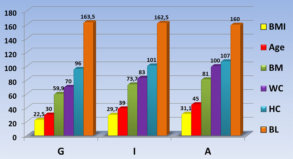
Given the above, groups with participants featuring different types of adipose tissue distribution had the biggest differences in the total body size when it came to the BM (G – 59.9 kg; I – 73.7 kg, A – 81.0 kg); WC (G – 70.0 cm, I – 83.0 cm, A – 100.0 cm), and age specifics – median age being 30 in G group, 39 in I group, and 45 in A group (Figure 1).

Figure 1. Variability of anthropometric parameters depending on the type of adipose tissue distribution